Author:Mingbo Wang
(Shenzhen Third People's Hospital Guangdong Shenzhen 518020)
[Abstract] Digital X-ray radiographic technology has a wide range of applications in clinical image evaluation. From traditional X-ray radiographic technology to today's digital X-ray radiography, the sharpness and accuracy of images have been greatly improved. In addition to digital X-ray radiography, dynamic DR radiographic technology has further broadened the thinking and practice of clinical application evaluation of X-ray radiography. Dynamic DR, through innovative technology fusion methods, enables X-ray radiography to break through the long-term imaging limitations of anatomical structures and enter motion functional imaging, providing important clinical value for imaging evaluation of diseases related to respiratory and joint systems.
[Keywords] dynamic DR; multi-function; motion function imaging; large view; dynamic video storage and playback
[Chinese Library Classification Number] R563
[Document Identification Code] A
[Article Number] 2096-3807(2020)10-0102-04
It can be said that digital X-ray radiography has experienced several generations of technological leap in the past ten years. With the development of digital flat-panel detector technology, in recent years, the direct digital flat-panel X-ray radiography system has developed into the most extensive digital radiography technology. The successful development of the digital flat-panel detector has greatly improved the clarity and accuracy of X-ray images, which is of great value for clinical applications. If say the first-generation digital radiography technology realizes image output through analog-to-digital conversion, then the second-generation digital imaging technology has changed the structure of traditional X-ray equipment through flat-panel detector. The traditional image chain consisting of image intensifiers, cameras, optical systems, and analog-to-digital converters is replaced by direct digitalization. It greatly reduces the complex chain that has many uncertain effects on image imaging, effectively reduces image noise and distortion, improves image contrast and resolution, and expands the dynamic range of the image by adjusting the window width and window level, and greatly improves the contrast and resolution of the image. The new third-generation dynamic digital X-ray radiography technology, based on the second-generation digital detector technology, breaks through the limitations of imaging technology, and has developed a dynamic digital detector based on the traditional static digital detector, the dynamic digital detector can achieve continuous multi-frame radiography, and through the collaboration of hardware, software, and image algorithms, output high-contrast and high-resolution dynamic images, which further broadens the scope, thinking and practice of clinical screening and diagnosis of X-ray radiography. This article focuses on the principle and clinical application of dynamic DR radiography technology to explore the future development direction.
1 Principles of dynamic DR radiography technology
The dynamic DR technology combines the radiation detection units into a flat linear array, which is directly connected to the large-scale integrated circuit, and completes the entire process of radiation reception, photoelectric conversion and digitization at the same time. Due to the direct conversion, many noise signals generated by transmission and signal conversion are reduced, and appropriate filter circuits are used to obtain low-noise and high-sensitivity images. Dynamic DR is composed of dynamic flat panel detector, high frequency generator, X-ray tube, intelligent motion system, computer, and image processing and transmission system. Compared with traditional digital X-ray radiography technology, Dynamic DR radiography can obtain multi-frame X-ray images with low-dose and high-speed within a time unit, after processing the system through the image algorithm, a continuous dynamic image (motion) is output at a high speed, realizing what you see is what you get.
Among them, the dynamic detector is the core component of the digital fluoroscopy X-ray machine, responsible for converting X-rays into digital image electrical signals, and then transmitting the image signals through the line, and then processing, displaying, filing and other operations to the image processing system. The dynamic flat-panel detector introduced here is an amorphous silicon X-ray flat-panel detector, which is an X-ray image detector with an amorphous silicon photodiode array as the core. The principle of static image imaging is that under X-ray irradiation, the CsI scintillator on the upper layer of the detector converts X-ray photons into visible light, and then the lower amorphous silicon array with photodiode function turns into image electrical signals, which are detected by peripheral circuits and A/D conversion to obtain digital images. The imaging process can be summarized as: X-ray - visible light - image electrical signal - digital image. The principle of dynamic image imaging is basically the same as that of static imaging, except that dynamic DR can be continuously shot at low dose and high speed and played continuously in real time on the computer, and finally displayed as a dynamic image.
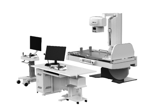
Angell Technology Talent Series - Remote Control Table Dynamic Flat-Panel DR
The dynamic flat-panel detector can be set in different working modes to realize a variety of different imaging frames, that is, continuous dot radiography at different frame rates and fluoroscopy functions at different frame rates. The realization process of dynamic imaging takes the pulse working mode as an example. The working mode of the detector and the parameters of the high-voltage generator are set by the software. When the exposure hand switch is pressed or the exposure foot switch is stepped on, the high-voltage generator host makes the tube emit X-rays, and outputs an exposure pulse signal to the detector, controls the detector to expose the image, and the image signal is transmitted to the computer via the transmission line , real-time display after software real-time processing, so as to realize dynamic imaging. The number of exposure pulses output to the detector per second is determined by the working mode of the detector. If the detector mode is 10f/s, the high-voltage generator will give the detector 10 exposure pulses per second. The dynamic detector is characterized by high conversion efficiency to X-ray, low energy loss, high signal-to-noise ratio, stable performance, and relatively small weight and volume.
2 Clinical applications and advantages of dynamic DR
Generally speaking, compared with CT, digital X-ray radiography has the characteristics and advantages of low dose, short examination time, high compliance, high image clarity, and low price. However, for general digital X-ray radiography, all two-dimensional images are obtained. At the same time, the image quality is difficult to control due to the influence of the subject, such as breathing and movement under examination. Compared with conventional digital X-ray radiography, dynamic DR can greatly improve the effect of X-ray image quality control. At the same time, it can provide a perspective and evaluation reference for motion functions for radiographic diagnosis of many parts, which can further improve the precision of screening and diagnosis. As an X-ray radiography technique, dynamic DR has a wide range of clinical applications and advantages, especially in imaging examinations of the chest, bones and joints, and abdomen. Compared with conventional static DR examinations, dynamic DR has unparalleled clinical applications Advantage.
Take the diagnosis of lung nodules on chest radiography as an example. Pulmonary nodules are a very common manifestation on chest imaging. Pulmonary nodules are small, focal, round, and imaging manifestations of increased density shadows, which can be single or multiple, without atelectasis, hilar enlargement, and pleural effusion. Solitary pulmonary nodules have no typical symptoms. They are usually single, clearly defined, dense, opaque X-ray shadows ≤ 30mm in diameter and surrounded by air-bearing lung tissue. Local lesions with a diameter> 30mm are called lung masses. There are two types of pulmonary nodules: multiple nodules and single solitary nodules. Most solitary nodules have insignificant clinical symptoms and are often found during chest X-ray examination. The chest has a good natural contrast, and X-ray examination is an important means to find and diagnose lung nodules. In the differential diagnosis of benign and malignant pulmonary nodules, a more accurate diagnosis can often be made by combining various signs of pulmonary nodule imaging and patient clinical data. Finding pulmonary nodules should be comprehensively analyzed from the location, nodule size, lesion shape, density, cavity, pleural changes, and hilar and mediastinal lymphadenopathy.
In recent years, with the popularization of DR and CT equipment, the number of lung nodules found in chest examinations has increased significantly. The accurate diagnosis of solitary lung nodules has always been a thorny problem in imaging diagnosis. Take the following picture as an example:
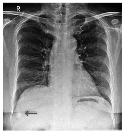
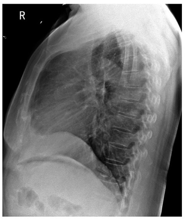
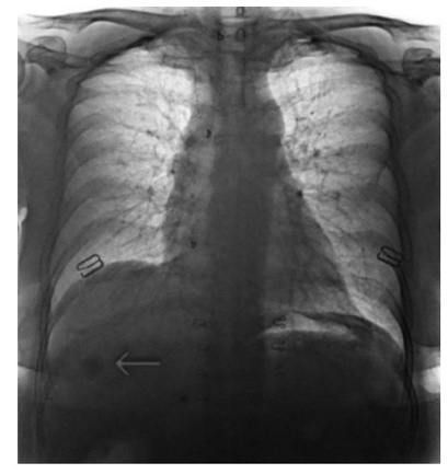
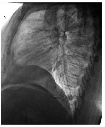
Female, 52 years old, with no symptoms. One month ago, a frontal chest radiograph was taken on a static flat DR in the outer hospital. The diagnosis report showed the shadow of the right subdiaphragmatic nodule. Considering the lesion of the intrahepatic nodule, Ultrasound examination of the liver is recommended. Ultrasound examination of the liver showed no abnormalities. Recently, a chest X-ray examination in our hospital revealed that there was a round high-density nodule with a diameter of about 17×15 mm under the right diaphragm, with clear edges. Immediately, the patient was observed with dynamic rotation and multi-position under fluoroscopy, and it was clear that the lesion was a nodule in the posterior basal segment of the right lower lobe, not a nodule in the liver (as indicated by the red arrow). CT examination is recommended. Three days later, a chest CT scan confirmed a round nodule in the posterior basal segment of the right lower lobe with calcification. CT diagnosis of induration in the posterior basal segment of the right lower lobe. The special feature of this case is that the right lower lobe nodule lesion is located in the posterior costophrenic angle area, and the position is lower and posterior. In the static DR, the chest was taken in the frontal position, and the nodular lesions were projected under the diaphragm. The static DR could not be visualized and rotated, and the lateral chest radiography was not taken. In addition, the examiner had insufficient experience, which caused misdiagnosis. This case is sufficient to prove the importance of dynamic DR visual fluoroscopy and the multi-angle observation function of rotation in clinical application, which can effectively avoid and reduce missed diagnosis and misdiagnosis.
|
|
CHEST PA: The shadow of the right lung nodule is projected under the diaphragm (←). | CHEST Right: The shadow of the nodule is located in the posterior basal segment of the right lower lobe, the posterior costophrenic angle area (→) |
|
|
|
Fluoroscopy single frame image 1: The shadow position of the right lung nodule during deep inhalation (←) | Fluoroscopy single frame image 2: The shadow position of the right lung nodule during deep inhalation (←) | Fluoroscopy single frame image 3: The shadow position of the right lung nodule during deep exhalation (←) |
Not only that, in the chest dynamic imaging analysis, whether it is DR, CT or MRI equipment, they are all static imaging. At the same time, CT and MRI equipment are both lying position imaging. In the examination of chest diseases, it is impossible to determine the pathogenic mechanism of the motion function of lung breathing. The functional motion imaging features of dynamic DR can technically realize the quantitative analysis and observation of the lung breathing process, so it would be more accurately diagnose of COPD and chronic obstructive pulmonary disease.
3. Application advantages of dynamic DR in COVID-19
The New Coronavirus (2019-nCOV) that broke out at the end of 2019, the main imaging findings of the patient are: bronchitis and bronchiolitis, localized patchy or mass shadows of pneumonia, and when the disease is severe, it appears as diffuse lungs of multiple consolidation shadows. Static DR chest radiography have a high rate of misdiagnosis and misdiagnosis in the initial stage of the disease screening. Compared with the static DR screening, dynamic DR can conduct a more comprehensive assessment of organ function and morphology through observation of dynamic images in the early stage, combined with imaging signs of CT. For example, by dynamically observing the dynamic changes of breathing to determine whether the diaphragm function is weak or other problems, it provides a more direct imaging basis for clinical treatment. In addition, the dynamic DR has the characteristics of remote control of the compartment, and at the same time, through functions such as one-key intelligent positioning and rotating foot pedals, it can avoid the contact between the operating doctor and the patient and reduce the risk of infection.
At the same time, for patients with COVID-19 during the recovery period, dynamic DR can more comprehensively and effectively evaluate the recovery of the patient's lung motion function by observing the dynamic process of the patient's lung breathing and the changes in lung ventilation.
4 Dynamic DR technology development direction
As the development and progress of X-ray radiography technology, dynamic DR has improved the accuracy of X-ray inspection. As a motion function imaging technology, dynamic DR will play a unique value in the analysis of lung and respiratory diseases such as COPD and wheezing. According to statistics from the World Health Organization, respiratory diseases are mainly represented by chronic obstructive pulmonary disease (COPD) and asthma, and are called the four major chronic non-communicable diseases together with cardiovascular and cerebrovascular diseases, cancer, and diabetes. The death toll from disease accounts for about 71% of the total death toll in the world. Due to the aging population, smoking and second-hand smoke, air pollution, the use of biofuels, and the large differences in the availability of medicines in different regions, the management of chronic respiratory diseases in my country is very severe. At present, the mortality rate of chronic respiratory diseases in China is 68 per 100,000, which has become the third leading cause of death from chronic diseases in China. There are about 100 million chronic obstructive pulmonary patients and about 30 million asthma patients in China, and the prevalence increases significantly with age. This not only brings a heavy economic burden to patients, their families and society, but also becomes the current major public health issues.
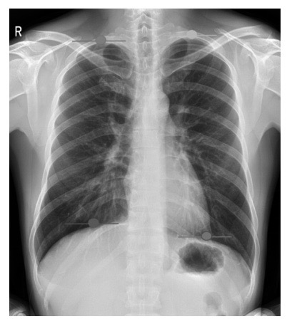
Quantitative trajectory of diaphragm movement
Current respiratory disease examinations mainly include: general pulmonary function examination, blood flow nuclear medicine examination, local pulmonary blood flow examination, and pulmonary thrombus location examination by enhanced CT. General pulmonary function tests cannot be used to diagnose changes in blood flow in the lungs, and enhanced CT is expensive. Dynamic DR technology can perform quantitative trajectory analysis on the movement of the diaphragm, which can effectively solve the evaluation and diagnosis of diaphragm adhesion and diaphragm function movement changes that have been unable to solve ordinary X-rays for decades. In addition, in the future, through the addition of AI intelligent algorithms, it can realize the quantitative change analysis of lung ventilation and blood flow with changes in lung density, quantitative comparative analysis of lung field area in the respiratory phase, and at the same time low cost, providing a more brand-new diagnostic idea for clinical diagnosis and practice.
5 Conclusion
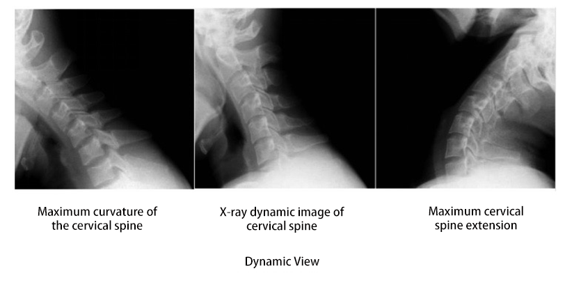
In summary, dynamic DR has the fusion and innovation characteristics of X-ray radiography technology. Its integration is mainly reflected in the realization of dynamic DR multifunctional application through technological fusion innovation, which can provide general medical solutions for imaging departments, targeting gastrointestinal, gastroenterology, orthopedics, gynecology, pediatrics, urology, emergency and routine DR radiography can provide corresponding solutions. The dynamic digital X-ray imaging system (Dynamic DR) has high-quality images and image post-processing. The imaging area is 17×17 inches. It can observe the dynamic images of the whole chest or digestive tract without moving the detector, and the images are clear, which is very helpful for doctors to quickly locate the lesion. Moreover, the core advantage of dynamic examination is that it can locate the target area under visual conditions, observe the activity and morphological changes of the lesion during deep breathing, and can rotate the patient's position for multi-angle observation to avoid the chest bone, heart shadow and hilar of the lesion, overlapping or covering large blood vessels leads to missed diagnosis and misdiagnosis. Especially the lung lesions located in the lateral sulcus on both sides of the chest and back spine, when the chest is irradiated, the lesions are easy to overlap or cover the mediastinal shadow and heart shadow, and the lung base lesions of the outer basal segment and posterior basal segment of the right lung lower lobe, due to the posterior and low position, some lesions are located in the posterior costophrenic angle area, and the lesions are covered by the diaphragm or the lesions are projected under the diaphragm when the frontal shot is illuminated, resulting in missed diagnosis or misdiagnosis as subphrenic intrahepatic lesions.
Therefore, the visualization function of dynamic DR can be combined with deep breathing exercises to dynamically observe the chest, ribs, mediastinum, heart shadow, diaphragm, and chest lesions, lung lesions, and mediastinal lesions in breathing motion from multiple angles when rotating the patient's position. Changes in the status can effectively avoid missed diagnosis and misdiagnosis caused by incomplete and incompact observation. And with the further development of AI technology, dynamic DR technology is expected to become a new diagnostic technology for clinical X-ray photography, providing a broader and more accurate evaluation value for clinical diagnosis.
[References]
[1] Jinghua Zhang. Digital Radiography Technology and Its Clinical Application Research [J]. Chinese Community Physician, 2019,35(02):135+137.
[2] Xiaopeng Lu. Application of Digital X-Ray DR Radiography Technology in Radiology Department [J]. China Health Industry, 2017, 14(14): 62-63.
[3] Qian Zhu. Application of Digital X-Ray DR Radiography Technology in Radiology Department [J]. Health Herald: Medical Edition, 2015, 36(2):265-265.
[4] Fen Guo. Discuss the Application Value of Digital Radiography Technology in Radiology Department [C]//2016 National Chronic Disease Diagnosis and Treatment Forum, 2016:137-138.
[5] Mingbo Wang. Application Value of Using Dynamic DR for Upper Gastrointestinal Angiography in The Risk Assessment of Gastrointestinal Bleeding [J]. Imaging Research and Medical Application, 2019:63-65.
[6] Xinsheng You. The Superior Value of Dynamic DR in The Diagnosis of Occult Fractures [J]. Imaging Research and Medical Application, 2019: 63-65.


