
Author: Qin Yan, director of Department of Radiology, the Fifth People's Hospital of Yichang City
Project background and significance
Pneumoconiosis is a systemic disease caused by long-term inhalation of productive dust and retention in lungs during occupation activities, which lead to diffuse fibrosis (cicatrices) of lung tissue. It is not only a serious threat to the life and health of workers, but also a huge economic loss to the country and individuals, affecting social stability.
Occupational pneumoconiosis refers to the following 13 kinds of pneumoconiosis in the classification and catalogue of occupational diseases in China, namely 1.Silicosis; 2.Coal worker's pneumoconiosis; 3.Graphitosis s; 4.Carbon black pneumoconiosis; 5. Asbestosis; 6.Talcosis; 7.Cement pneumoconiosis; 8.Mica pneumoconiosis; 9.Potter's pneumoconiosis; 10.Aluminosis; 11.Welder's pneumoconiosis; 12.Foundry's pneumoconiosis; 13.The other pneumoconiosis that can be diagnosed according to “the Diagnostic and Pathological Diagnosis Standard of Pneumoconiosis”.
Application and Practice
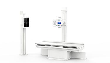
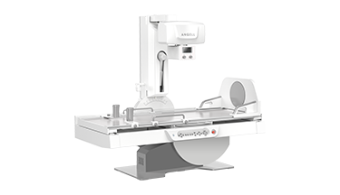
Multi-functional digital dynamic flat-panel DR is widely used in the clinical field because of its multi-function, high-definition imaging, real-time spot film, real-time playback and continuous spot film ability, radiography of all body parts and other functions.
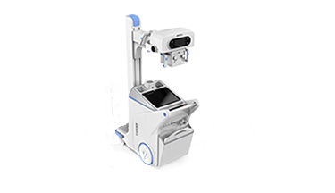
Our hospital is in Yiting District, the new industrial area of Yichang City, with thousands of dust-exposed workers. Our health examination center undertakes more than 95% of the tasks of occupational health examinations. After the introduction of dynamic flat DR (DTP570) of Angell Technology, more than 7000 chest radiography have been taken for various kinds of dust-exposed workers, and good results have been achieved.
Examination methods and parameters of dynamic DR for chest x-ray
The radiography position adopted in our hospital is posteroanterior (PA) position. Adjust the hands on the back of the waist and buttocks, arms adducted and shoulder joint internal rotation. Try to avoid the overlap of scapula and lung field. The center line of the x-ray tube is aligned with the level of the sixth thoracic vertebra, keep the position and take pictures after full inhalation and breath holding. Using fixed grid, The KV value is 90-125Kv; Mas value is 2-8Mas, which can be adjusted appropriately according to the body thickness of the patient.
Advantage analysis of dynamic DR examination
The following is the comparison between the High KV chest film taken by dynamic DR and the national pneumoconiosis diagnostic standard film in our hospital:
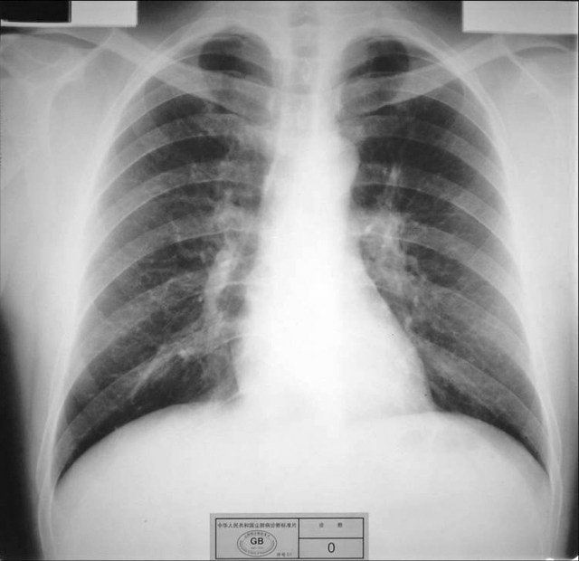
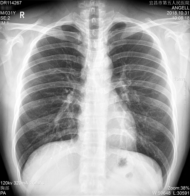
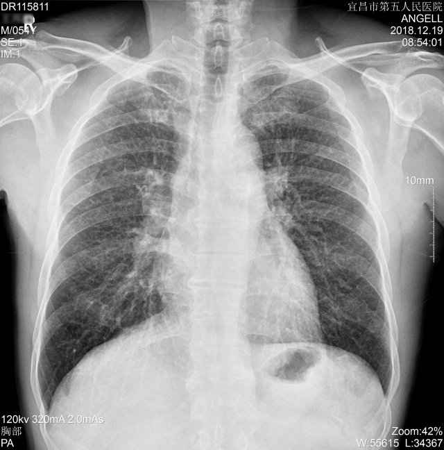
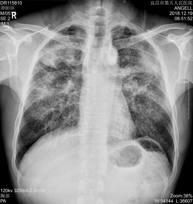
a. No pneumoconiosis: clear shadow of blood vessels in both lungs; normal chest image.
Standard chest image (Figure 1):
Chest image of dynamic DR (Figure 2):
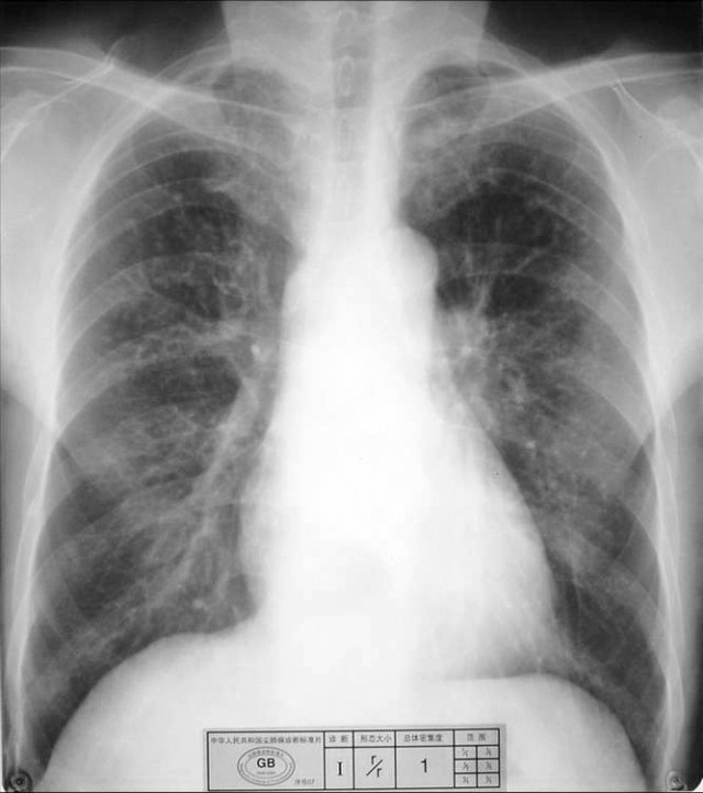
b. Phase I: the overall density of small shadow is level 1, and the distribution range reaches 4 lung areas
Standard chest image (Figure 3):
Chest image of dynamic DR (Figure 4):
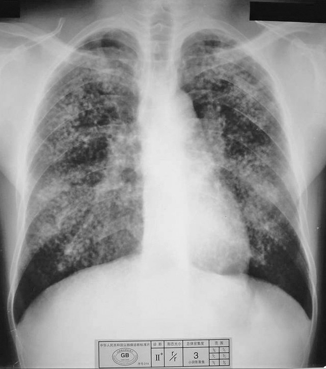
c. Phase III: the density of small shadow is 3 grade, and the distribution range is 6 lung areas
Standard chest image (Figure 5):
Chest image of dynamic DR (Figure 6):
Analysis
Dynamic DR has the characteristics of good image quality, high quality imaging rate and relatively low X-ray exposure. Through the image comparison, we found that dynamic DR can completely meet the requirements of pneumoconiosis diagnosis in terms of image clarity and X-ray penetration. And in the process of operation, we can switch to perspective mode at any time to identify the shadow, thus increasing the accuracy of the initial diagnosis. In conclusion, we believe that the application of dynamic DR in the examination of occupational pneumoconiosis is promising.