
Author: You Xinsheng | Zhangye Reproductive Medicine Hospital
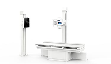
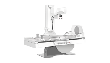
With the development of digital technology, digital medical imaging diagnostic equipment has evolved from analog digital imaging to current direct digital imaging. Its imaging speed, image quality, sharpness, contrast, and resolution has been significantly improved, greatly improving the accuracy of imaging diagnosis. The Dynamic DR combines multiple functions such as visual contrast, large-view multi-angle swivel HD fluoroscopy, HD radiography, millisecond-level radiography during fluoroscopy, real-time image saving and playback, automatic exposure control (AEC), whole body image stitching, and image post-processing. The aim is to get rid of many inconveniences of darkroom operation and to achieve a multi-purpose machine, making its clinical application scope and prospects wider.
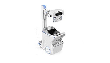
Compared with medical institutions at or above the secondary level, primary medical institutions are limited by funding, medical building construction area, and medical equipment configuration standards. Their medical diagnostic equipment is mostly basic equipment like conventional x-ray machines or Static DR and the inspection items and methods are simple. The diagnosis quality is not high, and the rate of missed diagnosis and misdiagnosis is increased.
In order to improve the accuracy of clinical diagnosis in primary medical institutions and to reduce and avoid misdiagnosis, the author summarizes the clinical application experience of our hospital in the past four years using ANGELL Dynamic DR as follows:
1. The visualization of contrast meets the routine contrast examination of clinical imaging in primary medical institutions.
Most of the radiographic examinations by basic medical institutions in their daily diagnosis and treatment are oral barium gastrointestinal contrast, barium enema contrast, oral or intravenous biliary tract, gallbladder contrast, intravenous pyelography, and hysterosalpingography. Under visualization conditions, these contrasts can not only reflect the shape, position, size, and contour of the organs, but also reflect changes in dynamic functions and the structure of mucosal folds in the lumen. For example, double contrast of the digestive lumen against the negative contrast agent (gas) clearly shows the application of barium sulfate on the mucosal surface, thereby showing the fine structure of mucosal folds. It has certain advantages for the discovery of early cancerous changes, small lesions and superficial lesions. It’s one of the test methods for early gastric cancer.
In oral or intravenous biliary tract contrast, cholecystography, and intravenous pyelography, Dynamic DR visualization can be used independently of the fixed time sequence of traditional methods. Dynamic DR fluoroscopy can be used to dynamically observe the biliary tract, gallbladder or pyelonephritis, ureter, and bladder. According to the diagnostic requirements and inspection purposes, it is possible to take large-full-view-abdominal radiography in real time to avoid information omission or scraps caused by blind radiography.
These examination methods are simple, easy, painless, low cost, easy to accept for patients, and have irreplaceable functions of CT, ultrasound, and gastroscopy in the examination and diagnosis.

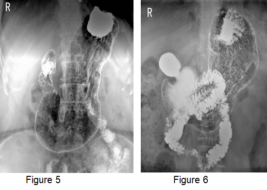
Figure 5, Figure 6 Barium forms a good daub on the surface of the gastric cavity, and the microstructure of the gastric wall and mucosal folds are clearly displayed.
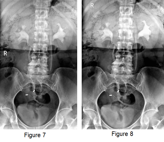
Figure 7, Figure 8 Intravenous pyelography, large-full-view abdominal.
Using Dynamic DR visualization to perform hysterosalpingography, you can intuitively understand the shape and size of the uterus, whether there are distortions, dilations, luminal adhesions and infarctions in each part of the fallopian tube. Under pressure bolus injection, the light and moderate blocking of fallopian tubes of patients can be unblocked, which plays an effective therapeutic role.
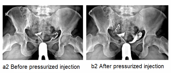
Blockage of the interstitial part of the right fallopian tube (Figure a2), which can be unobstructed after pressure bolus injection (Figure b2)
2. The large-view multi-angle swivel HD fluoroscopy helps to make the diagnosis of hidden rib fractures.
Hidden rib fractures are diseases that are difficult to find in time with conventional x-ray examinations and have a high rate of miss-diagnosis. The reason lies in the characteristics of the rib's physiological anatomy. The bone is curved in a bow shape, and it obliquely moves forward and downward from the posterior spine. The superior iliac ribs overlap with many tissues and organs, such as the mediastinum, heart, front and back rows of ribs, and pulmonary blood vessels. The inferior iliac ribs are affected by imaging factors of the abdominal cavity. They lack natural contrast and often have unclear images. The diagnosis of hidden rib fractures is difficult, and it is easy to miss. It needs to be combined with swivel fluoroscopy to accurately locate it. Some rib fractures are misdiagnosed and missed due to minor trauma and inconspicuous displacement of the fracture end, and insignificant indirect signs.
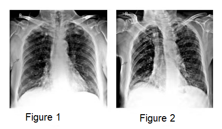
Figures 1 and 2 HD radiography images during fluoroscopy in deep breaths show axillary fracture of the 5th rib of the right chest, with a slight misalignment of the broken end.
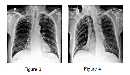
Figures 3 and 4 HD fluoroscopy and radiography images during fluoroscopy in deep breaths show the fracture of the 5th rib fracture of the right chest is reduced, and the fracture line is unclear.
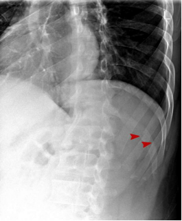
Figure 5 Dynamic DR swivel multi-angle fluoroscopy and radiography clearly shows the fracture line.
When the ribs are incompletely fractured or the displacement of the complete fractures is not obvious, it can only appear as a slight uplift, depression, folds or incomplete translucent line shadows and dense shadow. It sometimes even only appears as an abnormal curvature of the ribs without visible fracture lines. Therefore, some extremely fine fracture lines and incomplete fractures must be seen combined with HD radiography and dynamic swivel fluoroscopy, multi-angle and multi-position observation.
The reasonable application of dynamic DR large-view multi-angle swivel HD fluoroscopy can effectively avoid missed diagnosis and misdiagnosis.
3. HD fluoroscopy with radiography significantly improves the accuracy of heart and lung disease diagnosis.
During the examination of heart and large blood vessel diseases, the Dynamic DR HD fluoroscopy function is applied, and the video saving function is selected at the same time, collecting dynamic video images of different positions of the heart and large blood vessels. You can also see barium swallowing if it is necessary. According to the image information of the heart and blood vessels obtained from different positions you can observe the changes in the position, shape and size of the heart and large blood vessels, as well as the amplitude, intensity, rhythm of the heart and large blood vessels. You can see whether there is displacement of the opposite beat points, atrioventricular notches, and interventricular grooves, or if there is deepening and displacement of esophageal pressure marks. It can be determined which room or chamber is enlarged, and the extent of the increase, and the decrease or increase in pulmonary blood flow on both sides, etc., can be correctly judged in combination with clinical symptoms and signs.
Compared with the conventional X-ray machine and Static DR, the multi-functional combination of Dynamic DR significantly improves the image quality, image sharpness, and resolution, and avoids the loss of lesion information and scraps caused by blind radiography. Swivel the patient's position at will under visualization conditions, which facilitates the observation of multiple positions and angles of the lesion, and obtains more information about the lesion, avoiding the omission of lesions and lesion characteristics caused by single position radiography. It is especially beneficial to find lesions that overlap with mediastinum, heart shadow, thoracic bones, and sub condylar ribs. It is widely used in lung cancer examination.
4. Real-time image saving and playback to promote clinical teaching.
The Dynamic DR image real-time saving and playback function has been widely used in clinical consultation and teaching, which greatly improves the accuracy of disease diagnosis.
The multi-functional clinical application of Dynamic DR provides a reliable diagnostic basis for the diagnosis and treatment of diseases and plays an indispensable role in primary medical institutions.