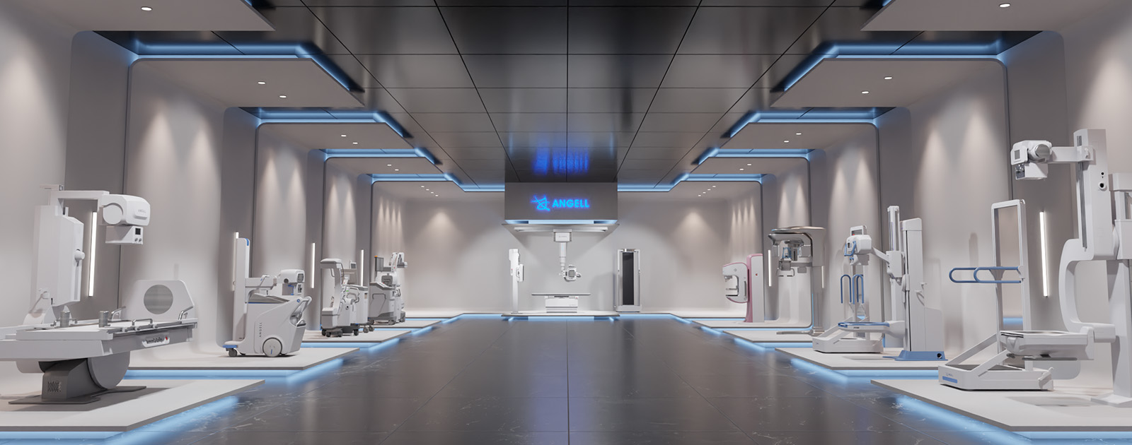
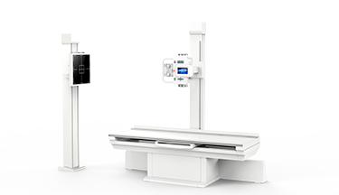
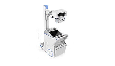
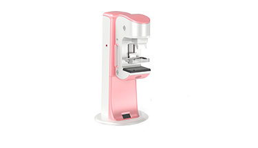
The 84th China International Medical Equipment Fair kicked off in Shanghai on May 13, with leading manufacturers from home and abroad showing up at the fair. In the exhibition area of medical imaging, there are various new products, and all leading manufacturers have brought their own innovative technologies and products. Among which, Angell Technology, a leading manufacturer in the field of general radiology, released the first weight-bearing 3D scanning solution based on dynamic DR system in China: WR-3D (weight-bearing dynamic 3D image reconstruction system), realizing the true three-dimensionality of general radiological equipment, leading the general radiological equipment from 2D imaging to 3D imaging in an all-round way, and providing weight-bearing 3D accurate imaging through complementary CT, which has set off a new round of technological revolution in the radiology industry.
“Weight-bearing” is the translation term for weight-bearing, which generally means the natural weight-bearing position or standing position. First, let’s look at a set of clinical weight-bearing 3D images reconstructed by Angell Technology’s WR-3D technology.
How does Angell Technology’s WR-3D technology based on dynamic DR system realize the weight-bearing 3D image reconstruction? And what is the clinical significance of this technology? Let’s find out.
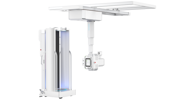 Figure: WR-3D
I. Orthostatic 3D image reconstruction technology based on dynamic DR system.
WR-3D is a weight-bearing 3D image reconstruction system, which has a number of patents for invention. Different from the conventional CT reconstruction system in which the plate and the tube rotate synchronously, it adopts the industry’s pioneering way of rotating the foot pedal to drive the patient to obtain the weight-bearing 3D scanning data, which retains the original shape and operation process of DR equipment. It adopts a semi-open and encircling space capsule design, with a 360-degree rotating and movable platform, surrounded by a waterfall of science fiction light strips, bringing a comfortable and technological experience during examination. Large-format automatic rotation scanning technology achieves a maximum FOV of 350mm, and supports scanning of both legs and bilateral hip joints. The full 3D cone beam reconstruction algorithm has no cone angle artifacts and guarantees voxel isotropy. Scanning in 30 seconds, and drawing in 2 minutes.
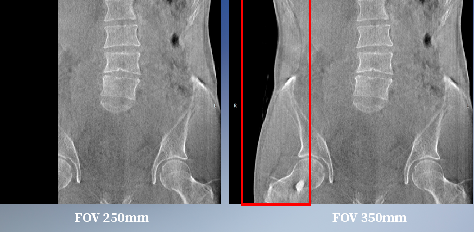
Figure: FOV 350mm at maximum
II. Systemic 3D scanning and reconstruction in weight-bearing state.
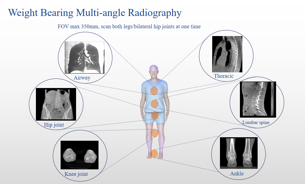
Figure: Systemic scanning
Angell Technology’s WR-3D system enables 3D scanning and reconstruction of the entire body, including airway, cervical spine, lumbar spine, hip joint, knee joint and ankle, in the natural weight-bearing position. In addition, the system can reconstruct weight-bearing tomographic images, MPR images, MIP images, and VR volume rendering images, and perform distance and angle measurements, so as to facilitate accurate preoperative planning and accurate postoperative evaluation.
III. WR-3D technology leads general radiology to the era of 3D accurate diagnosis in an all-round way, and provides weight-bearing 3D images through complementary CT.
With the advent of WR-3D technology, it has mainly solved two core problems: 1. It has solved the current situation of high rate of missed DR diagnosis in general radiology. Conventional DR equipment has 2D images at a certain angle only and poor density information. According to relevant statistics, due to factors such as placement and tissue overlap, the misdiagnosis and missed diagnosis rate of 2D plain film is as high as 20% or more, while WR-3D can obtain richer diagnostic information, take 3D reconstruction images from multiple angles, and obtain the information of arbitrary angles and sections as well as highly sensitive density information. 2. It has solved the problem of CT’s inability to obtain orthostatic (weight-bearing) 3D images. The non-weight-bearing images of conventional spiral CT cannot reflect the joint disease information in weight-bearing state. For clinical diagnosis, especially in orthopedics, the weight-bearing images are of more clinical value. The muscle state, joint space and skeletal force line of the patient in weight-bearing state are significantly different from those when the patient is lying flat. Therefore, WR-3D has a very important clinical value in accurate preoperative planning and accurate postoperative evaluation.
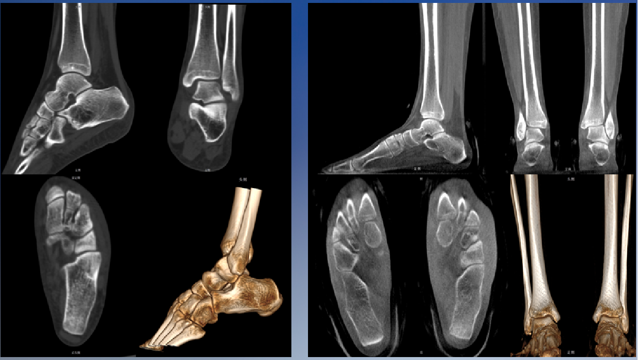
Figure: Providing weight-bearing 3D images through complementary CT
In general, WR-3D technology is the true 3D technology applied to DR equipment. Under the premise of not changing the original shape and operation process of DR equipment, WR-3D technology can not only enrich the diagnostic information of DR in general radiology to improve the diagnostic accuracy, but also well make up for the function deficiency of CT’s inability to obtain weight-bearing 3D reconstruction images.
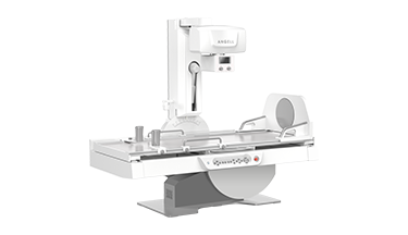
At this CMEF, Angell Technology also released a full line of general radiological products. Among which, as one of the world’s two largest true 3D suspension DRs, the “Zhumu” suspension dynamic DR, a popular suspension dynamic DR, is eye-catching.
The “Zhumu” suspension dynamic DR integrates the latest technology of current dynamic imaging technology. It is an efficient and intelligent suspension dynamic DR in routine X-ray radiography, an efficient and accurate suspension dynamic DR in dynamic radiography such as overlapping lesions and occult lesions, and an efficient and convenient suspension dynamic DR in comprehensive clinical applications such as systemic stitching, angiography and pneumoconiosis examination. Meanwhile, “Zhumu” is also one of the world’s two suspension dynamic DR products that support the weight-bearing 3D image reconstruction function, leading the three-dimensional development of general radiological equipment.
I. Dynamic imaging and complementary CT.
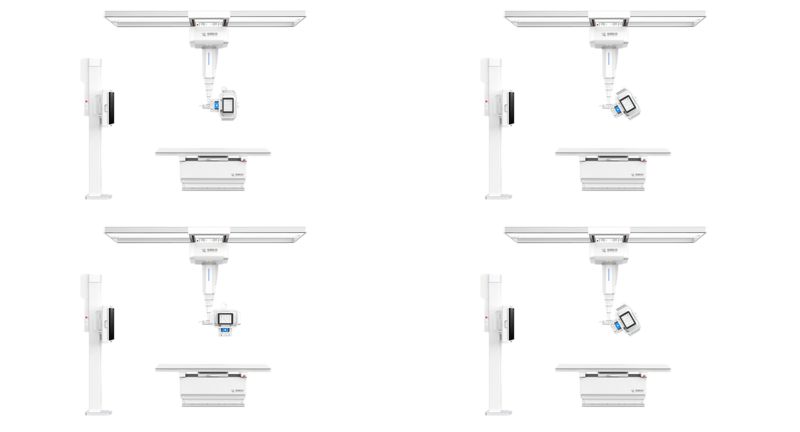
Zhumu Figure 1
Zhumu is equipped with one dynamic detector and one static detector independently developed by Angell Technology. The dynamic flat panel detector has a large format size of 17*17 inches, 9.4 million high-definition pixels, and 0.8S extremely fast dynamic and static high-definition spot film function. A number of precise diagnostic functions, such as dynamic observation combined with 0.8S extremely fast dynamic and static switching spot film, dynamic local magnification, etc., can help doctors accurately capture lesions. Moreover, “Zhumu” supports 3D tomography and reconstruction of the patient’s scanned site in weight-bearing state, and complementary CT.
II. 4D ten-axis, efficient filming.
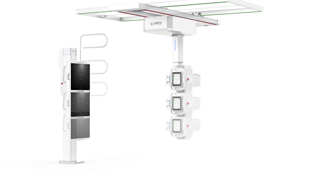
Zhumu Figure 2
Zhumu is equipped with a 4D ten-axis linkage motion system. It is the first suspension DR in the world to realize two-way automatic accurate tracking, and realizes the tube tracking flat panel and flat panel tracking tube in all three motion dimensions of orthostatic lifting, clinostatic lifting and clinostatic level. The tube and detector can be moved to a stroke of about 20cm close to the ground, allowing the filming range to cover the whole body conveniently. Meanwhile, a series of clinical practical designs, such as electric lifting bed, large intelligent touch screen, one-key switching between orthostatic and clinostatic positions, built-in voice intercom, and automatic exposure control by AEC, make all-position, multi-angle conventional X-ray radiography efficient and convenient.
III. Comprehensive application and orthostatic 3D.
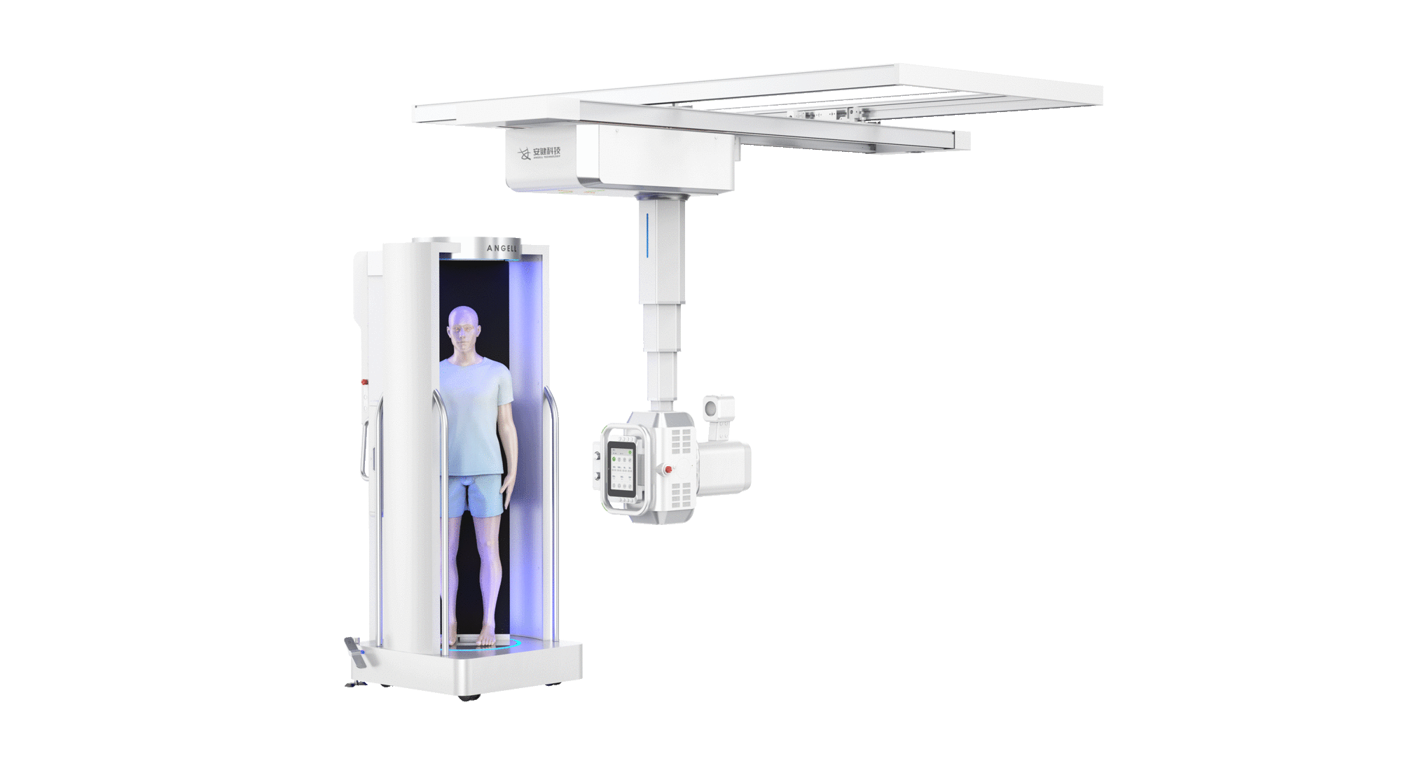
Zhumu Figure 3
“Zhumu” fully supports the functions of automatic long bone stitching, esophagography and professional pneumoconiosis examination. In addition, Zhumu is the only two suspension dynamic DRs in the world that can support weight-bearing 3D tomography and reconstruction, which solves the problem of orthostatic 3D imaging that cannot be realized by CT\NMR equipment, and supports multiple reconstruction functions such as tomographic image reconstruction, MPR multi-plane reconstruction, MIP maximum density projection reconstruction, VR volume rendering, etc.
Digital X-ray radiographic technology has ushered in a new technological revolution. Based on the characteristics of high efficiency and low cost of digital X-ray radiography, it can be predicted that 3D digital X-ray radiographic technology will usher in rapid development and become another diagnostic tool for hospitals. At present, the international giant Siemens and the domestic giant Angell Technology have already taken the lead. We look forward to this technology benefiting many more patients.

