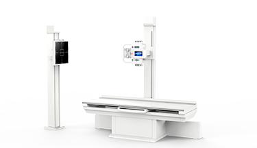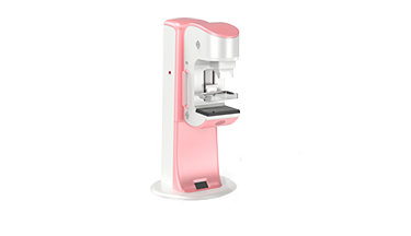

As the most widely used medical imaging examination in the world, X-ray radiography has covered the disease screening of billions of people. Since Roentgen discovered X-ray and widely used in medical fields, among the various X-ray technologies, general X-ray radiography including X-ray radiography, mammography, gastrointestinal and other X-ray examination, has become the most widely used examination in the world. With its convenience, low cost, low dose and high efficiency, X-ray radiography not only has unique clinical application value in chest, lung, bone and joint disease examination, but also has incomparable clinical advantages in breast disease examination. X-ray radiography has experienced rapid development in China. From the early analog imaging stage, indirect digital X-ray imaging stage, direct digital X-ray imaging stage to dynamic multi-function imaging stage, the general X-ray radiography technology has developed rapidly.
From the current technology perspective, where is the development direction of digital X-ray radiography? This paper summarizes the six trends of the rapid development of general X-ray technology by combing the technology exploration path of the current global digital X-ray industry chain.
Trend 1: Dynamic
At the RSNA 2019, Dynamic Digital Radiography Technology won the best innovation award of the American Society of radiologists. Generally speaking, compared with CT, digital radiography has the advantages of low dose, short examination time, high compliance, high image definition and low cost. But for the general digital X-ray radiography, it is difficult to control the image quality because of the influence of the object, such as breathing and movement. Compared with conventional X-ray, Dynamic DR can greatly improve the quality control of X-ray image. At the same time, it can provide evaluation reference for dynamic imaging diagnosis of many parts, and further improve the diagnosis accuracy. As an X-ray radiography technology, Dynamic DR has a wide clinical application value and advantages. For example, Dynamic DR has the visualization function in chest imaging diagnosis, which can exclude the overlapping or covering of chest skeleton, heart shadow and pulmonary hilar vessels when rotating the body position at will, combined with respiratory movement. Multi angle dynamic observation of the chest, ribs, mediastinum, heart shadow, diaphragm, as well as chest lesions, lung lesions, and mediastinum lesions in the state of respiratory movement can also meet the clinical needs in the prompt of various vessel positions and the evaluation of treatment effect. At present, the trend of dynamic digital X-ray radiography technology has become a common understanding in the industry. It can be predicted that with the further development of Dynamic Digital X-ray Technology, it is expected to completely replace conventional static radiography, fundamentally change the clinical disadvantages of missed diagnosis of general digital X-ray radiography, and greatly improve the accuracy of DR screening.
Trend 2: Three Dimensional
The second trend of digital X-ray radiography technology: Three-Dimensional development. SIEMENS Healthineers releases new high-end suspended 3D digital X-ray Multitom Rax in 2020, which is called "LI", can realize 3D scanning of bone and joint and reconstruct 3D digital image through surrounding motion of double suspended x-ray tube manipulator and detector manipulator, especially for the parts that need 3D inspection under weight-bearing position, including knee joint, ankle joint and other parts. In addition to SIEMENS, ANGELL TECHNOLOGY, a leading global manufacturer of digital X-ray radiography device, also innovates the path of digital three-dimensional technology, realizing three-dimensional scanning of bone and joint through 360 degree automatic rotation scanning device. Through the clinical research of 3D digital X-ray of SIEMENS and ANGELL TECHNOLOGY, we can see that 3D digital X-ray scanning can realize the 3D image information in the weight-bearing position, and solve the lack of 3D image information in the standing position that cannot be solved by CT and MRI. It has high clinical application value for the diagnosis of orthopedics, and has been recognized by many international experts. With the breakthrough of this technology, the future of general digital X-ray radiography is expected to be further combined with three-dimensional technology. Through general DR, many parts of the two-dimensional screening and three-dimensional examination can be carried out, fundamentally changing the disadvantages of high missed diagnosis rate of digital X-ray radiography. At the same time, the high efficiency and low cost of digital X-ray examination will further promote the rapid development of three-dimensional digital X-ray technology.

Left: SIEMENS 3D scanning suspension system; Right: ANGELL 3D scanning suspension system
Trend 3: Functionalization
The third trend of general digital radiography: functionalization. For a long time, doctors have a strong demand for the assessment of dynamic status of body tissues and organs. General digital X-ray radiography, including CT tomography, cannot directly present dynamic imaging, such as the motion imaging of chest, cervical and limb. Through the assessment of dynamic imaging, we can accurately evaluate the disease before and after diagnosis and treatment. At present, dynamic imaging technology has played a unique clinical diagnostic value in the application of chest motion imaging, such as COPD, asthma, cervical spondylosis and other related diseases. Taking COPD as an example, we can accurately evaluate the pulmonary motion stage by comparing the changes of COPD respiratory lung field area and normal respiratory lung field area.

Evaluation of pulmonary motion of COPD patients
In addition, motion function assessment can also accurately assess the disease information that causes respiratory motion insufficiency, severe clinical symptoms and mild lung image, such as extensive pleural adhesion, pericardial effusion, chronic bronchitis, emphysema, diaphragmatic paralysis, etc. When such clinical manifestations are found in time, the comparison of inspiratory and expiratory images or dynamic imaging can provide corresponding information when the clinical manifestation is found in time.
Trend 4: Spectroscopy
The fourth trend of general digital radiography: Spectroscopy. The X-ray absorption coefficient of any substance can be transformed into the absorption coefficient of any two base substances and reached the same level as that of the substance X-ray attenuation effect by energy spectrum X-ray. Therefore, it can transform the attenuation of a substance into the density of two substances producing the same attenuation. According to the absorption coefficient of a certain base substance energy level, the density and spatial distribution of the base substance can be evaluated, so as to realize the preliminary analysis of substance composition and substance separation, and produce the image of biomass separation. Energy spectrum technology can achieve ultra-low-dose photography at the same time, obtain high-quality image. In addition, the information of energy spectrum curve, material separation image and effective atomic number map can be achieved by energy spectrum technology, which provides a more intuitive and accurate method for the analysis of lesion components. The development of digital X-ray spectroscopy will make the digital X-ray photography a revolution development and at present it has made great achievements in the laboratory stage.
Trend 5: Ultra-Low Dose Quantification
The fifth trend of general digital radiography: ultra-low dose quantification. For a long time, digital X-ray radiography has been pursuing the optimization between the optimal image and the optimal dose. At the same time, there is an extensive discussion about the relationship between the appropriate dose and the optimal image. For general digital X-ray photography technology, ultra-low dose quantification has always been the direction of global efforts and a number of medical imaging research teams are committed to the realization of ultra-low dose digital X-ray imaging around the world. At present, we can see that with the further improvement of detector working efficiency and the continuous optimization of work flow and post-processing algorithm, the radiography dose is gradually decreasing. In addition, with the further development of detector technology, ultra-low-dose X-ray radiography is expected to make a greater breakthrough. The R&D team of Angell Technology has been publishing two research articles in the top journal, Nano Energy and Nanotechnology. The research results show that: the silicon-based heterojunction perovskite quantum dot nanocrystalline array directly converts X-ray photon signals into current signals. It effectively avoids the process of converting X-ray photons into visible light and then converting visible light into electron-hole pairs and improves the efficiency of quantum detection. Because the perovskite materials contain Pb and Br atoms with high atomic numbers, the thickness of the perovskite array can be achieved in millimeters, ensuring strong X-ray absorption.

The latest research results of detectors based on perovskite materials
In addition, several scientific teams including the Shenzhen Institutes of Advanced Technology (SIAT) of the Chinese Academy of Science (CAS), Southeast University, University of Science and Technology of China, Jilin University, etc. have also made many great achievements in the research of perovskite materials, laying a favorable foundation for the technical application of perovskite. It is believed that with the breakthrough of related technology, the ultra-low dose quantification of digital X-ray radiography will be widely used.
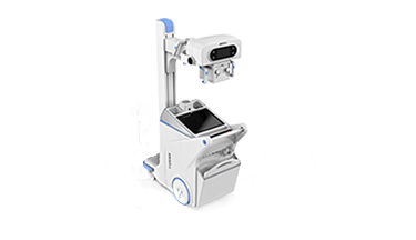
Trend 6: Portability
The sixth trend of general digital radiography: portability. In recent years, with the rapid development of mobile DR, portable X-ray device has been widely used in clinic. Different from the bulky and large fixed digital X-ray device, portable X-ray device can flexibly work in various clinical departments. With more efficient and simple examination workflow, mobile X-ray photography can be carried out for inconvenient circumstances anytime and anywhere. During the COVID-19, mobile digital X-ray device has become the main medical device for daily imaging follow-up. It plays an important role in fighting against global epidemic. In addition,with the acceleration of various emergency use, including the demand for first aid and emergency treatment under various natural disasters, light portable X-ray devices are developing rapidly all over the world.
To sum up, after a hundred years of development, general digital X-ray device still has a broad technical and application development. Due to the unique advantages of general digital X-ray device, it always has irreplaceable value in clinical application. With the dynamic technology, multifunctional development and three-dimensional development, the practical value of general digital X-ray device in clinical diagnosis and treatment will be further redefined. It can be expected that in the era of precision medical diagnosis and treatment, general digital X-ray device will make a new practice, making the general radiology work with other medical imaging technologies to better benefit human health.