
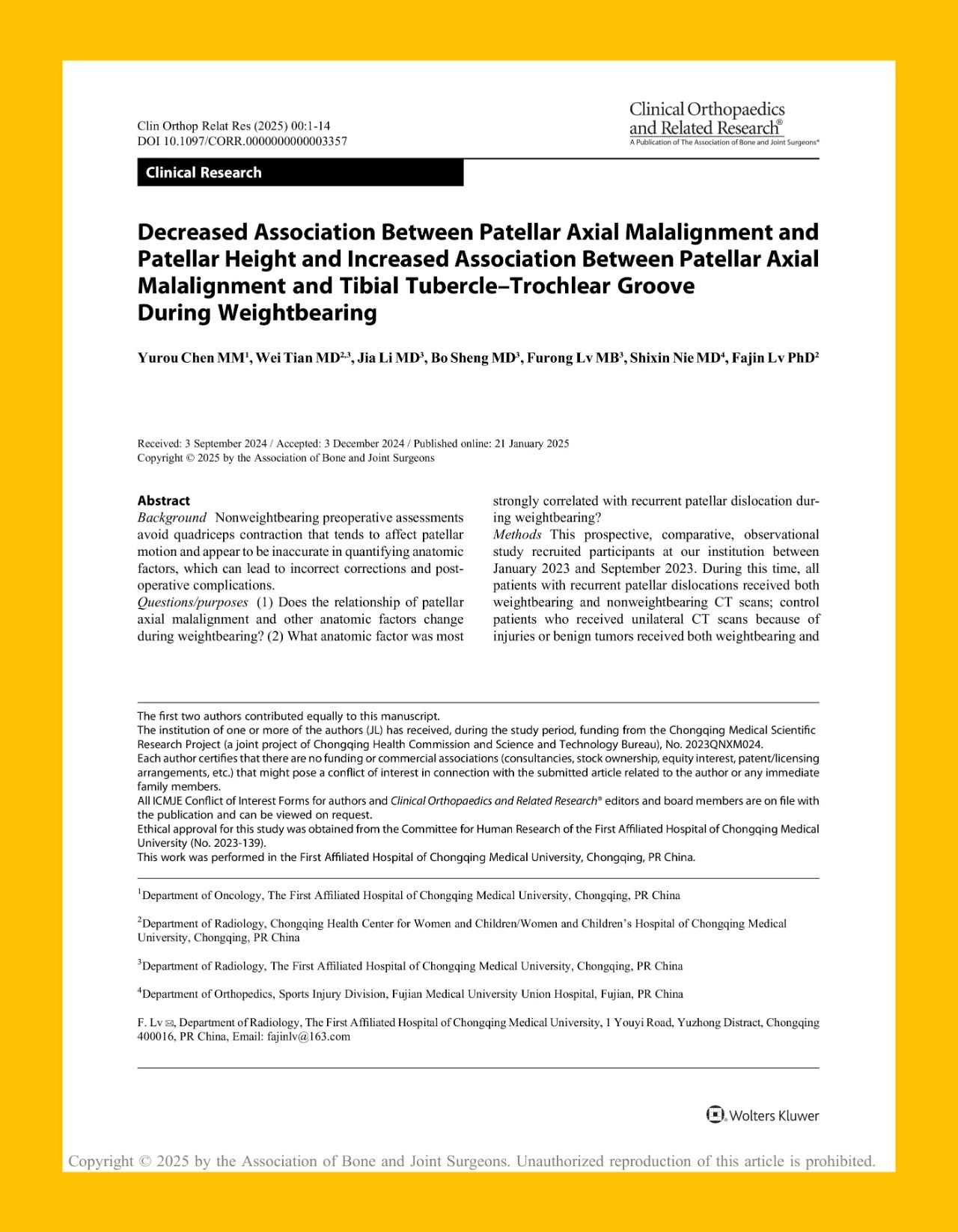
Chongqing, China – A research team from the First Affiliated Hospital of Chongqing Medical University has published a groundbreaking study in Clinical Orthopaedics and Related Research® (CORR), highlighting the diagnostic superiority of weightbearing cone beam computed tomography (CBCT) in evaluating patellar stability. The study, titled “Decreased Association Between Patellar Axial Malalignment and Patellar Height and Increased Association Between Patellar Axial Malalignment and Tibial Tubercle-Trochlear Groove During Weightbearing”, provides compelling evidence that WR-3D weightbearing CBCT significantly improves the accuracy of patellar malalignment assessment compared to traditional imaging methods.
Challenges in Diagnosing Recurrent Patellar Dislocation
Recurrent patellar dislocation (RPD) is a common yet complex condition in sports medicine and orthopaedics, posing diagnostic and therapeutic challenges. Early identification of anatomical abnormalities and dynamic instability is critical, yet current diagnostic approaches are limited by the complexity of etiology, lack of standardized surgical criteria, and insufficiently individualized postoperative rehabilitation.
Traditional imaging techniques—including radiographs, CT, and MRI—are predominantly performed in a supine, non-weightbearing position. X-rays assess parameters such as trochlear depth (>145° indicating dysplasia), congruence angle (CA), and patellar height (Caton-Deschamps Index >1.2). CT quantifies tibial tubercle–trochlear groove (TT-TG) distance (>20 mm is considered abnormal) to determine the need for tibial tubercle osteotomy. MRI enables detailed evaluation of medial patellofemoral ligament (MPFL) tears—present in approximately 90% of acute dislocations—as well as cartilage and bone bruising. However, none of these methods accurately reflect the patellofemoral joint’s biomechanical status under load.
Innovative WR-3D Technology Yields Breakthrough Insights
In collaboration with Angell Technology’s DTP580-B3 (WR-3D) weightbearing CBCT platform, the research team conducted a clinical study that directly addresses the limitations of conventional non-weightbearing imaging. The study found that WR-3D enables more accurate visualization of the correlation between patellar axial malalignment and TT-TG distance under physiological load, reflecting real-time muscular engagement, particularly of the quadriceps.
This innovation provides a major leap forward in patellar instability diagnostics, allowing clinicians to observe the patella in a functional, weightbearing state—offering a more comprehensive and physiologically relevant understanding than traditional supine imaging.
Significance of Weightbearing Imaging
The study also reveals that patellar positioning and alignment under weightbearing conditions differ significantly from those in a relaxed, supine position. This underscores the clinical importance of evaluating patients in a functional stance to better predict instability and dislocation risk. By simulating real-life joint loading conditions, WR-3D overcomes the limitations of static supine assessments and facilitates biomechanically informed decision-making.
About the Journal and Editorial Endorsement
The findings were published in Clinical Orthopaedics and Related Research® (CORR), a leading journal in orthopaedic medicine published by The Association of Bone and Joint Surgeons®. Founded in 1953, CORR covers a wide array of topics including joint surgery, sports medicine, trauma, musculoskeletal oncology, and biomechanics. The journal maintains a strong academic reputation, with a 2023 impact factor of 4.2 and a Q1 ranking in both the Orthopaedics and Surgery categories according to JCR metrics.
Study Design and Methodology
The research included 52 patients with recurrent patellar dislocation and 52 healthy controls, all examined between January and September 2023. Participants underwent both weightbearing CBCT and traditional CT scans. During WR-3D scanning, patients stood upright on an automated rotating platform with full knee extension and even weight distribution.
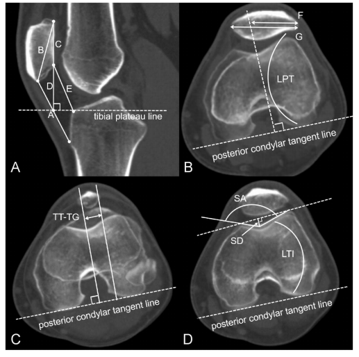
Key anatomical parameters were evaluated, including patellar height, axial malalignment (assessed via bisect offset index and tilt angle), TT-TG distance, and trochlear morphology (depth and sulcus angle). All measurements were independently conducted by two experienced radiologists with a minimum four-week interval between assessments to ensure reliability.
Key Findings
The study revealed a markedly stronger correlation between patellar axial malalignment and TT-TG distance in weightbearing CBCT compared to non-weightbearing CT. These results confirm the diagnostic superiority of WR-3D in capturing the true biomechanical behavior of the patellofemoral joint under functional conditions.
By reflecting quadriceps activation and realistic loading, WR-3D overcomes the diagnostic shortcomings of traditional static imaging and sets a new standard for evaluating patellar instability.
Future Outlook and Clinical Impact
This research is part of a series of clinical innovations made possible by Angell Technology’s first-generation WR-3D. Building upon this foundation, Angell has continued to refine weightbearing CBCT solutions—enhancing scanning range, imaging performance, and automatic measurements.
Angell’s DTP580-B3 (WR-3D) is the world’s first weightbearing cone beam CT capable of full-spine and lower-limb 3D scanning and tomographic reconstruction in a standing position. It is also the only solution globally that integrates automatic analysis across the entire musculoskeletal chain—including the spine, pelvis, hip, knee, and foot & ankle—bridging diagnostics and treatment planning.
In just two years, nearly 100 WR-3D systems have been installed across China, driving adoption and clinical integration of weightbearing CBCT. Through expanded collaboration with clinical institutions, Angell aims to further broaden the clinical value and global impact of WR-3D technology, enabling truly personalized orthopaedic care.