WR-3D medical imaging system

WR-3D

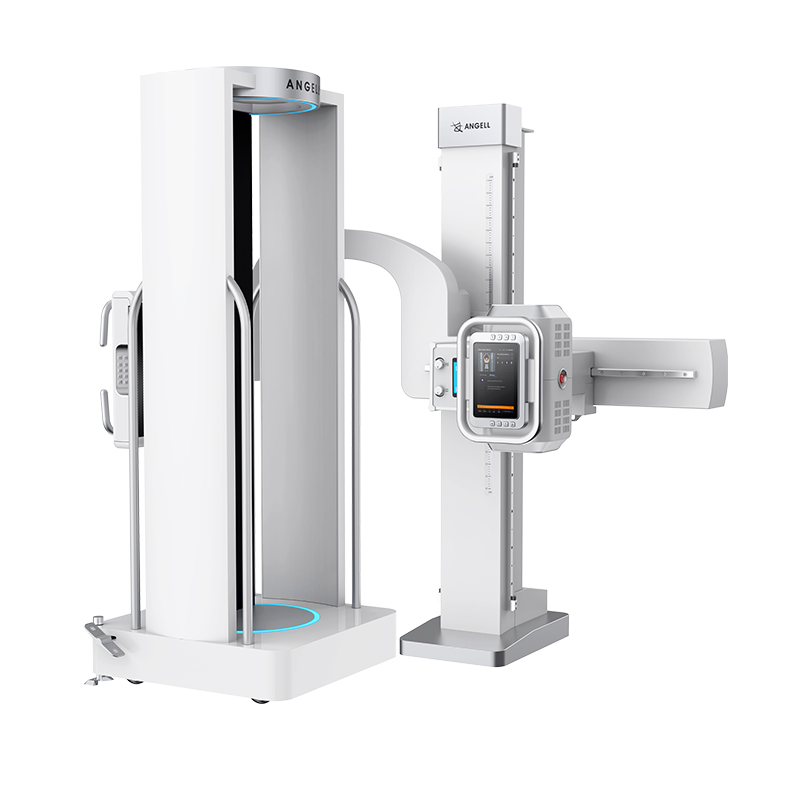
WR-3D is Angell Technology's weight-bearing radiographic 3D imaging system for spine, lower limb, hip, knee, and ankle assessment, combining volumetric reconstruction, AI measurement, and 2D fluoroscopy support.

Weight-Bearing Radiographic 3D system

Product Detail

 1.   Complete 3D scanning

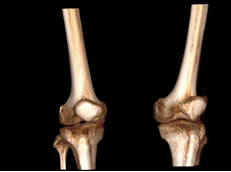
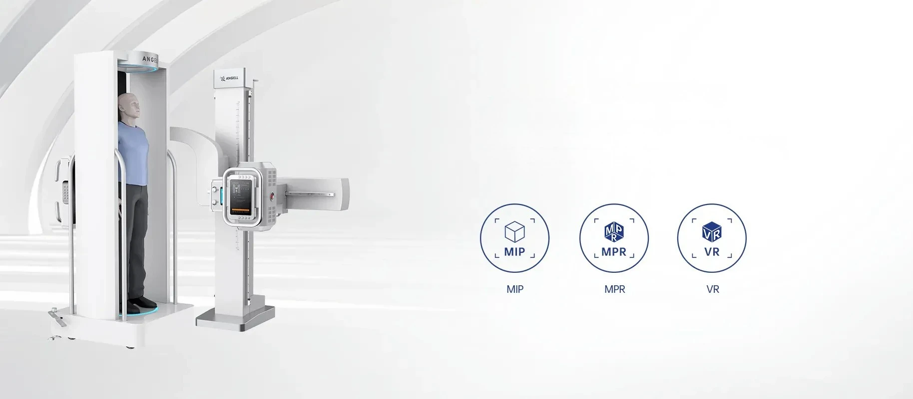
WR-3D utilizes volumetric imaging technology to achieve full three-dimensional reconstruction of the scanned area in a weight-bearing position. It delivers comprehensive views across coronal, axial, and sagittal planes, and supports reconstruction of MIP, MPR and VR.

2.   Full Spine/Full Lower Limbs Applications

WR-3D supports comprehensive 3D scanning of the full spine, full lower limbs, and specific joint regions including the hip, knee, and ankle.

3.   Fully automatic AI measurement

WR-3D provides AI-assisted measurements for scoliosis and knee osteoarthritis assessment, supporting orthopedic planning and follow-up review.

4.  2D+Fluoroscopy

WR-3D combines weight-bearing 3D imaging with 2D radiography, stitching, fluoroscopy and contrast study workflows in one platform.

WR-3D weight-bearing radiographic 3D system for full spine scanning and measurement

World's First Weight-Bearing Full Spine Scanning and Measurement System

WR-3D complete 3D scanning with MIP MPR and VR reconstruction

Complete 3D scanning

WR-3D reconstructs weight-bearing 3D datasets into coronal, axial and sagittal views, with MIP, MPR and VR modes for reviewing the scanned anatomy.
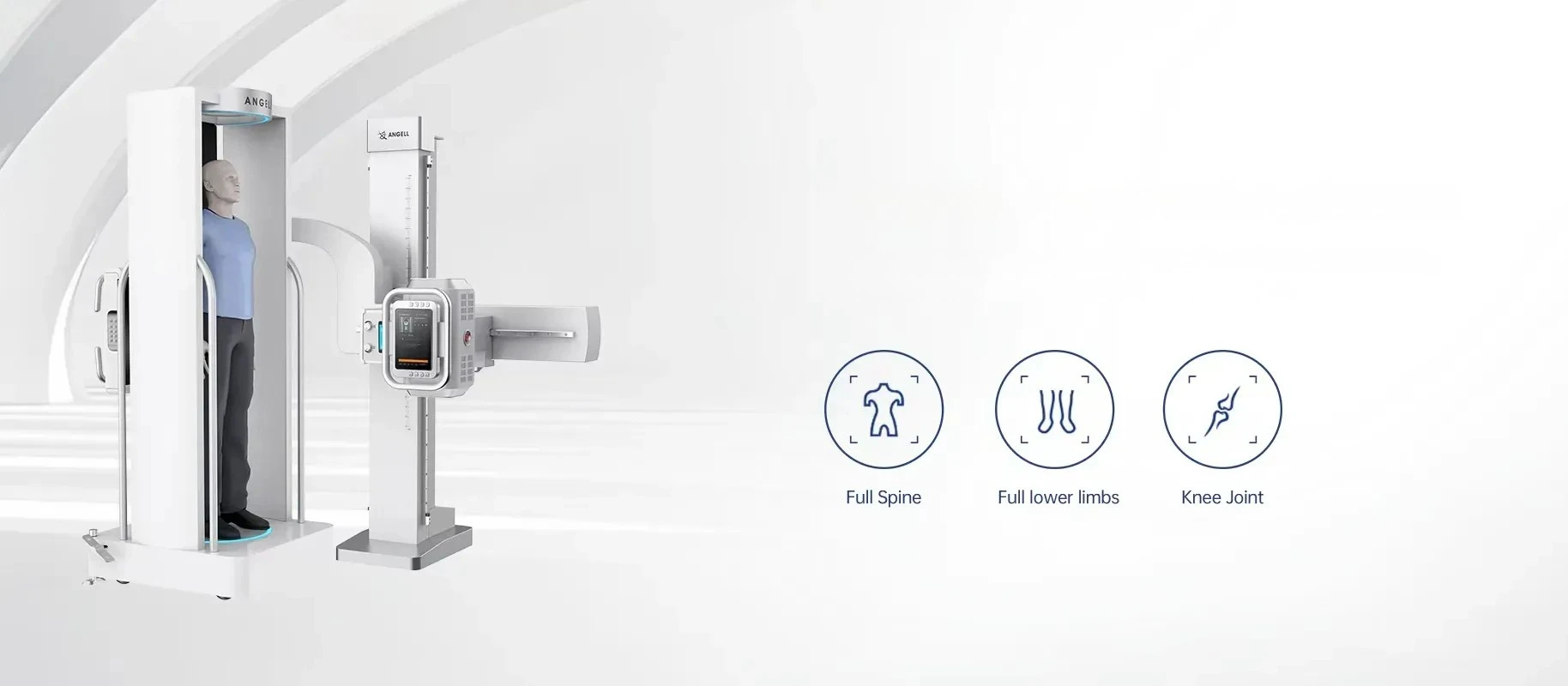
WR-3D full spine full lower limb and knee joint applications

Full Spine/Full Lower Limbs Applications

WR-3D supports standing 3D review of the full spine, full lower limbs and key joint regions including the hip, knee and ankle.

WR-3D fully automatic AI measurement for scoliosis and knee osteoarthritis assessment

Fully automatic AI measurement

WR-3D provides AI-assisted measurements for scoliosis and knee osteoarthritis assessment, supporting orthopedic planning and follow-up review.
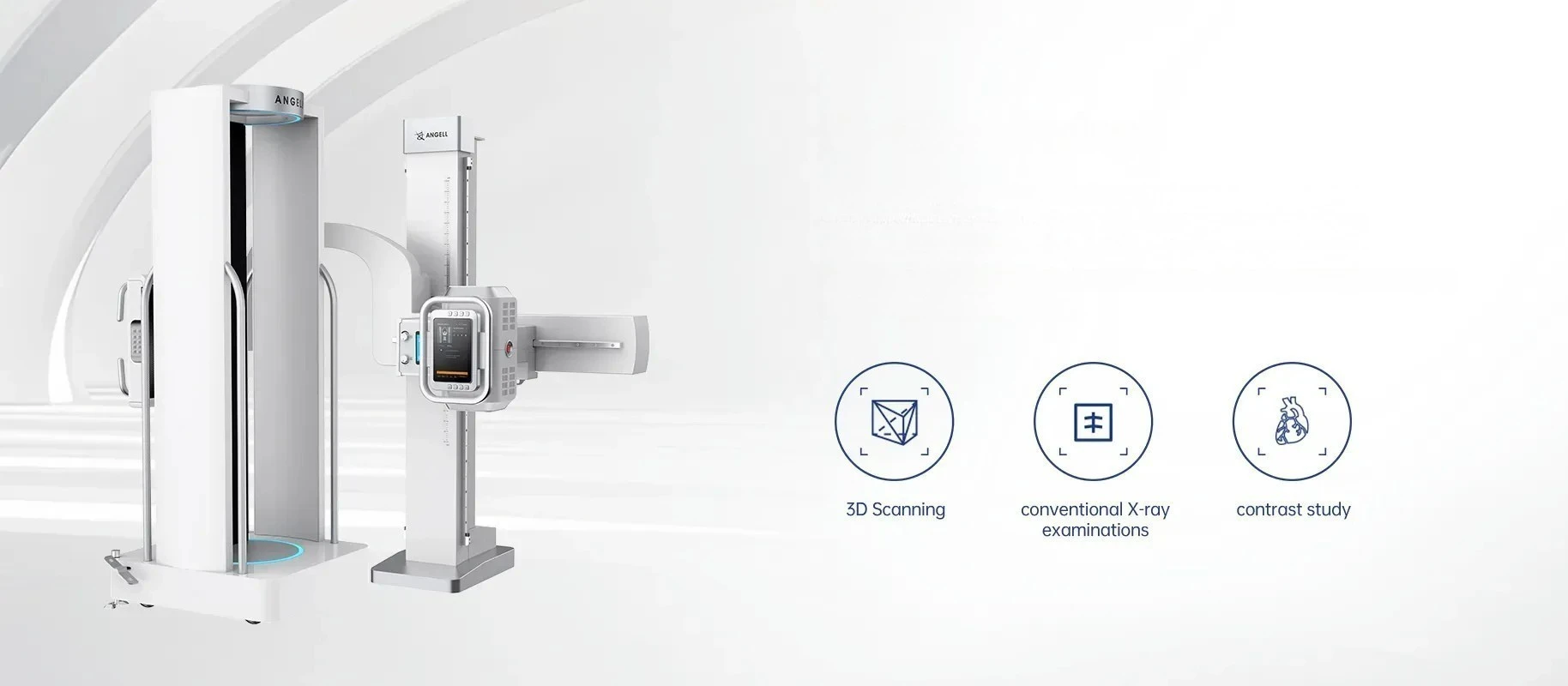
WR-3D 2D X-ray fluoroscopy and 3D scanning workflow

2D+Fluoroscopy

WR-3D combines weight-bearing 3D imaging with 2D radiography, stitching, fluoroscopy and contrast study workflows in one platform.

3D Imaging Demo

See how WR-3D presents reconstructed 3D views

This video demonstrates the WR-3D 3D imaging display, showing how reconstructed volumetric views can support review of skeletal anatomy beyond a single flat projection.

Standing 3D scan visualization Spine, lower-limb and joint review context Orthopedic weight-bearing workflow

Standing cone-beam acquisition

Fixed imaging chain for standing weight-bearing acquisition

WR-3D uses a fixed X-ray tube and flat-panel detector while the patient stands on a motorized platform between them. The workflow is built for weight-bearing orthopedic assessment, where posture control during rotation is part of image quality, not a separate setup detail.

1

Defined scan geometry

The tube-detector relationship stays fixed while the patient platform rotates through the acquisition path.

2

Weight-bearing posture control

Patient instruction and stance confirmation are part of acquisition setup because weight shift can affect standing 3D reconstruction.

3

Motion-aware workflow

Controlled platform rotation keeps posture consistency within the scan workflow instead of treating rotation as only a timing metric.
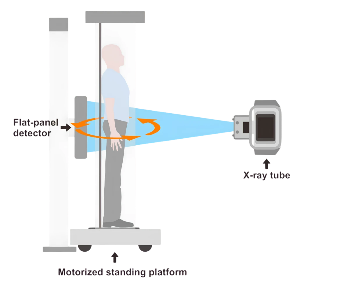
WR-3D standing cone-beam acquisition diagram with flat-panel detector, X-ray tube and motorized standing platform
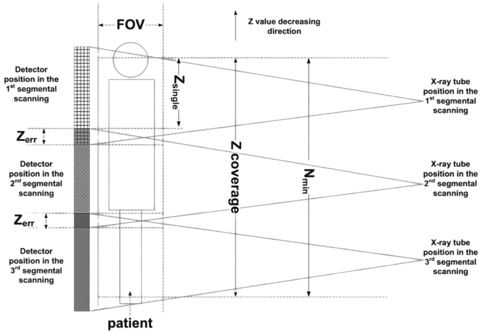
WR-3D multi-sequence full-spine scanning and Z-axis stitching coverage diagram

Full-spine 3D stitching

Multi-sequence scanning extends Z-axis coverage

For long anatomical regions, WR-3D acquires successive standing scan segments along the vertical axis. The reconstructed segments are combined into long-field views for spine or lower-limb review while the patient remains in a weight-bearing position.

1

Standing segment acquisition

Each segment is captured with the patient upright, avoiding a supine-to-standing posture transfer.

2

Z-axis extension

Successive scan ranges are used when the clinical question requires extended spine or lower-limb coverage.

3

Protocol-based output

Stitched views are generated within the selected reconstruction protocol, keeping extended review tied to the scan setup used for that examination.

Orthopedic assessment in WB posture

Weight-bearing views for spine, lower-limb and joint review

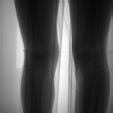
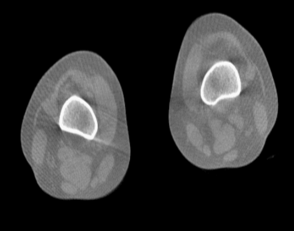
WR-3D is positioned for orthopedic scenarios where standing posture changes anatomical relationships, including scoliosis assessment, lower-limb alignment, knee joint review and postoperative follow-up. Reconstructed slices and 3D views help separate structures that overlap in a single projection.
Get A Quote