
At Angell, we start every X-ray vs CT vs MRI comparison with the same question: what information does the examination need to deliver? X-ray provides projection views, CT separates anatomy into cross-sectional slices, and MRI distinguishes soft tissues through differences in their magnetic properties. Throughout this guide, “X-ray” refers to conventional radiography; CT also uses X-rays.
For an imaging department, image detail is only half the picture. The examination might call for a standing view, full-length coverage, three-dimensional data under load, or imaging at the bedside. These practical requirements should guide equipment selection.
X-ray vs CT vs MRI at a Glance
These are different imaging capabilities, not steps in a mandatory sequence. Modality selection should follow examination-specific guidance such as the ACR Appropriateness Criteria, rather than an assumption that every workup must progress from X-ray to CT to MRI.
How X-ray, CT, and MRI Differ by Tissue, Depth, and Position
X-ray records a two-dimensional projection, CT separates anatomy into cross-sectional slices and volumetric reconstructions, and MRI differentiates tissues through signal contrast. Start with the information gap: is the missing piece a projection and alignment view, depth and geometry, or tissue characterization?
X-ray: Projection, Alignment, and Position-Dependent Views
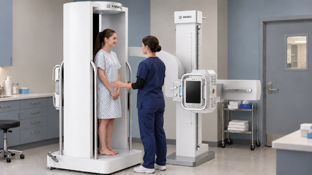
Radiography is the right tool when the clinical answer can come from an outline, density pattern, relative position, or alignment. The system configuration determines whether upright, standing, seated, recumbent, stretcher, or bedside positions are available.
The trade-off is superimposition: anatomy along the X-ray beam is superimposed in the same projection. Before comparing systems, define the required projections, whether weight-bearing information matters, the anatomical length to cover, and the patient’s ability to hold each position.
A department that needs both standing and recumbent views should test both workflows on the same unit. Check detector reach, tube-detector alignment, field coverage, and patient access. DTP580, for example, supports projections such as Waters, patella axial, calcaneal axial, and cross-table lateral views. When overlap prevents localization or depth assessment, CT becomes the next consideration.
CT: Cross-Sectional Localization and 3D Relationships
CT earns its place when the unanswered question is about depth or three-dimensional relationships. The scanner reconstructs multiple X-ray projections into slices that can be reviewed in different planes and rendered as three-dimensional models.
With appropriate protocols, CT can depict bone, soft tissue, and blood vessels, although MRI may separate some soft-tissue types more clearly. CT removes projection overlap and provides spatial localization, while radiography preserves a direct view acquired in a specified position.
When evaluating CT equipment, define the required coverage, reconstruction planes, contrast workflow, and review location. Conventional CT is generally table-based, so patient transfer, table access, motion control, and acquisition position need confirmation. A weight-bearing radiographic 3D system follows a different pathway from conventional table-based CT.
MRI: Soft-Tissue Differentiation and Sequence-Dependent Contrast
Choose MRI when the decision hinges on distinguishing muscles, tendons, ligaments, nerves, cartilage, bone marrow, or other soft tissues. The request should specify the anatomical region, required contrast information, and sequence set.
MRI avoids ionizing radiation, but motion sensitivity, scan duration, safety screening, coil selection, and patient tolerance shape the workflow. A regional MRI examination should not be assumed to deliver the full-length standing alignment information available from a purpose-configured radiographic workflow.
These modalities work together. Radiography can document alignment in the required position, CT can resolve depth and complex spatial geometry, and MRI can characterize tissues that projection or attenuation data alone may not separate.
Where X-ray Acquisition Changes the Information Available
Once requirements point toward X-ray-based imaging, review coverage, loading position, and acquisition location as three distinct requirements.
Full-Length 2D Coverage Without Image Stitching
When an examination assesses hip-to-ankle alignment, all required landmarks must be captured in the same full-length projection. The Changfeng Series uses a proprietary 47-inch large-format detector for full-spine and full-lower-limb imaging in a single exposure, without image stitching. It also offers automatic angular stitching for full-body recumbent imaging, a separate option for a different positioning need.
Standing Volumetric Imaging Under Load
A standing projection shows alignment under body weight, but overlapping anatomy remains. When the examination needs both a loaded position and a volume that can be reviewed from multiple angles, the acquisition requirement shifts. The WR-3D weight-bearing radiographic 3D system provides volumetric imaging for the full spine, full lower limbs, and hip, knee, and ankle regions. Reconstructions deliver slices in three planes and 3D visualization. With the corresponding AI option, it supports AI-assisted measurements for scoliosis and knee osteoarthritis assessment.
As a cone-beam CT system, WR-3D combines volumetric information with weight-bearing positioning. It also incorporates conventional radiography, full-length image stitching, and fluoroscopy for a mixed examination list. A 2023 validation study of extensive-coverage, weight-bearing cone-beam CT included 30 volunteers with adolescent idiopathic scoliosis and reported generally good clinical image quality. Under that protocol, the reported CTDIvol was 1.23 mGy.
Bedside Dynamic Imaging When Transfer Is Difficult
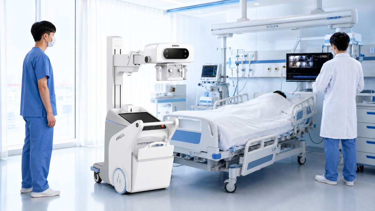
At the bedside, access becomes part of the imaging requirement. The Lingxi Series mobile dynamic digital radiography system brings high-resolution imaging and dynamic observation to emergency and bedside workflows, including placement and verification of PICCs and endotracheal tubes.
Dynamic observation adds a time dimension. Fluoroscopy displays successive X-ray frames to follow movement or guide a procedure. A request for portable X-ray should specify whether the department needs still images, dynamic observation, or both.
For a bedside configuration, the site survey should trace the system’s path from the corridor to the examination position: doorways, bedside clearance, detector placement, and space around monitoring equipment. The review should also track preparation, safety controls, image transfer, and reporting.
Compare Safety, Throughput, and the End-to-End Examination Workflow
Measure Time to a Reviewable Answer
Break the workflow into preparation, patient transfer, positioning, contrast administration when required, acquisition, reconstruction, and image transfer. CT typically acquires data faster than MRI, while a radiographic examination may involve one or several projections. The meaningful endpoint is when the required views reach the reporting team.
Patient position can reshape that pathway. A bedside radiographic system brings acquisition to the patient, whereas CT and MRI normally require access to a dedicated scanner. During a demonstration, record setup, acquisition, reconstruction, and transfer times separately.
Verify Modality-Specific Safety Controls
Radiography and CT use ionizing radiation. Verify the intended protocol, anatomical coverage, collimation, exposure control, repeat criteria, and dose record for each examination. MRI does not use ionizing radiation, but staff must screen people, implants, devices, and objects and verify MR safety conditions.
Test the Handoff from Acquisition to Reporting
Follow one representative examination from patient arrival through image review. Confirm that the required projections or reconstructions reach PACS or the reporting workstation, and document room readiness, calibration, training, support, and configuration-dependent outputs such as MPR, VR, segmentation models, and measurement reports.
FAQs About X-ray, CT, and MRI
Why Might an X-ray Still Be Needed After an MRI?
The two examinations may answer different questions. An MRI focused on a joint’s soft tissues does not necessarily include a full-length standing alignment view. Compare purpose, coverage, and patient position before treating the second study as a duplicate.
Does Every CT Scan or MRI Scan Require Contrast?
No. Contrast follows the examination protocol and the information required. Many studies are performed without it; others use it to make particular tissues or vessels easier to assess.
Can Dynamic X-ray Imaging Replace CT or MRI?
Not as a general replacement. A dynamic projection sequence adds information about movement or device position, but it does not produce a CT volume or MRI tissue contrast on its own.
How to Choose Between X-ray, CT, and MRI for Your Examination Mix
Equipment planning should start with the missing capability. Radiography fits when the answer is available from the correct projection, patient position, and anatomical coverage. CT fits when depth or three-dimensional geometry remains unresolved. MRI fits when the information gap concerns soft-tissue characterization.
Once the modality is defined, test one representative examination from positioning through acquisition, transfer, and review. For X-ray-based configurations, reproduce the intended full-length, weight-bearing, or bedside workflow rather than running a generic demonstration.
Send us your examination mix, expected volume, patient-positioning requirements, room dimensions or bedside access constraints, existing imaging equipment, and destination market. Our technical team can discuss system configurations, installation requirements, and application support. Contact us to discuss your X-ray imaging workflow.