
A mammography machine is a dedicated breast X-ray imaging system used for mammography. The RadiologyInfo mammography overview describes mammography as specialized breast imaging that uses a low-dose X-ray system to see inside the breasts.
The main difference lies in acquisition and output: FFDM, or full-field digital mammography, produces 2D projection images, while digital breast tomosynthesis, or DBT, captures multiple angled projections and reconstructs them into a scrollable image stack.
Mammography Machine Components: Quick Preview
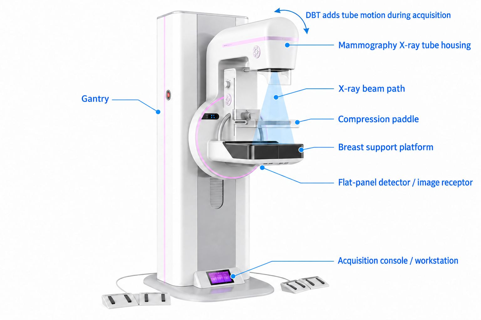
The mammography system turns an X-ray exposure into image data through several connected components.
Component | Role in the imaging chain |
|---|---|
X-ray tube | The imaging process starts here, with the tube producing the X-ray beam. |
Compression paddle | Keeps the breast steady during exposure. |
Detector | After the X-rays pass through the breast, the detector receives the remaining signal. |
Image-processing software | Detector data are then processed into a 2D image or reconstructed slices. |
Workstation | The finished images are shown here for review. |
What Does a Mammography Machine Do and When Is It Used?
A mammography machine creates low-dose X-ray images of breast tissue. It is used for screening when no breast symptoms are known and for diagnostic evaluation when a symptom or an abnormal screening result needs closer review.
During an examination, the breast rests on the detector and is briefly compressed before the system captures the X-ray signal. The FDA’s mammography guide notes that compression keeps the breast still and spreads the tissue more evenly, helping reduce overlap and produce a clearer image.
The same imaging method can be used for different clinical purposes. Screening mammography is performed when no breast symptoms are known and focuses on early changes that may not yet be felt, such as a small mass, asymmetry, or tiny calcium deposits. Diagnostic mammography is used when a specific concern has already been identified, such as a new lump, focal pain, nipple discharge, skin or nipple changes, or an abnormal screening result.
These examinations are commonly performed in hospital radiology departments, breast imaging centers, screening centers, and breast clinics.
FFDM vs. DBT: How Image Acquisition and Output Differ
FFDM and DBT differ mainly in how they acquire X-ray data and present breast tissue. FFDM captures a single projection for each view and produces one flat 2D image, so tissue at different depths may overlap. DBT captures multiple low-dose projections as the X-ray tube moves through an arc, then reconstructs them into thin slices for layer-by-layer review. The FDA’s explanation of digital mammography describes this multi-angle acquisition and slice reconstruction process.
Feature | FFDM | DBT |
|---|---|---|
Image acquisition | One 2D image from a single angle | Multiple low-dose images from different angles |
X-ray tube | Stays in one position during exposure | Moves through an arc |
Image output | One flat 2D image | A series of reconstructed slices |
Tissue overlap | Tissue at different depths appears together | The slices allow overlapping tissue to be reviewed layer by layer |
Image review | One flat image | Layer-by-layer review |
Technical Boundaries of DBT
DBT shows the breast as a series of reconstructed layers. Unlike CT, it views the breast from a limited range of angles instead of scanning all the way around. Different systems may use different scan angles, numbers of projection images, tube movements, and reconstruction methods. Exposure settings and movement during the scan can also affect the final images.
A 2D/DBT Imaging Workflow with the Fanghua Series
At Angell Technology, our Fanghua Series digital mammography system can support both FFDM and DBT in the same breast imaging workflow. After the breast is positioned and compressed, a standard FFDM view keeps the X-ray tube at a fixed angle and captures one projection with the detector, producing a flat 2D image.
When a DBT view is needed, the breast remains in position while the X-ray tube moves through an arc and captures multiple low-dose projections from different angles. The system reconstructs these projections into thin slices, allowing the radiologist to review the breast layer by layer rather than as a single flat image.
The system’s large-format detector is designed to cover the breast, lymph-node, and axillary areas. During acquisition, automatic exposure control helps set the exposure, while motorized collimation limits the imaging field and smart light-field adjustment helps align the area being captured. This workflow shows how the Fanghua Series can produce either a conventional FFDM projection or a reconstructed DBT image stack, depending on the imaging mode selected.
Frequently Asked Questions
Is every digital mammography machine a 3D mammography machine?
No. Some digital mammography machines support only 2D FFDM, while others include DBT or both 2D and DBT modes. “Digital” does not automatically mean “3D.”
Does a 3D mammogram also produce a 2D image?
DBT primarily produces reconstructed breast slices from multiple angled projections. Depending on the system, it may also generate a synthetic 2D image from the same DBT data. This software-generated image is different from a separately acquired FFDM image, which comes from its own 2D exposure.
Is DBT radiation dose always higher than FFDM?
Dose comparisons only make sense after the protocol and system are identified. The result changes with separately acquired 2D images, synthetic 2D images, exposure settings, equipment design, and local requirements.
Conclusion
FFDM presents the breast as a single flat projection, while DBT captures images from multiple angles and reconstructs them into slices for layer-by-layer review. At Angell Technology, we bring these two imaging approaches together in our Fanghua Series and broader digital mammography portfolio. Explore our digital mammography systems to see how we support conventional 2D and DBT imaging workflows.