
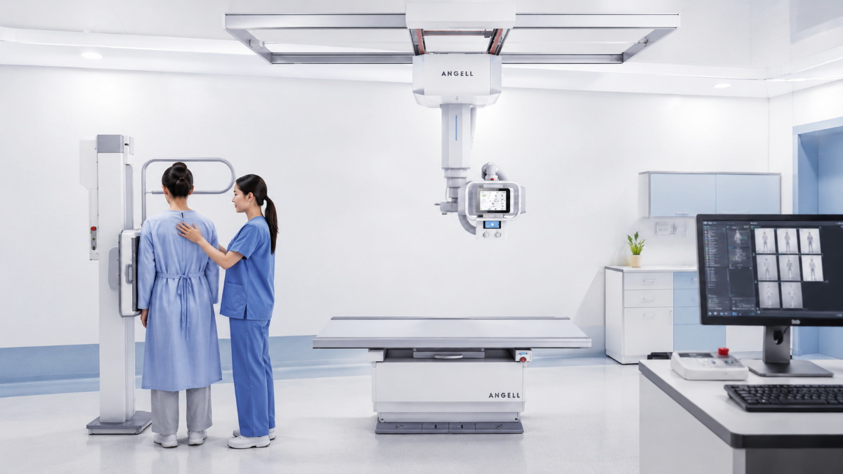
Choosing between dynamic DR and static DR should begin with the examination. Static DR is usually the right fit when one well-positioned radiograph answers the clinical question. Dynamic DR earns its place when movement, live positioning, contrast passage, device travel, or functional change affects how the exam is performed or interpreted.
When our technical team reviews a project, we start with three questions: What must the image show? Can the patient reach the imaging room safely? What workflow can the department support every day? Those answers are more useful than comparing detector size or generator output in isolation.
What Actually Changes When DR Becomes Dynamic?
Static DR produces one projection image per exposure. That is what routine chest, abdominal, spinal, and extremity examinations often require. When positioning is predictable, and a single view shows the anatomy adequately, dynamic acquisition may add complexity without changing the answer.
Dynamic DR adds time. It records a sequence that can show breathing, swallowing, joint movement, catheter travel, contrast passage, or alignment before a spot image is saved. The difference is therefore larger than “a photo versus a video.”
Dynamic capability still does not make every examination clearer or easier. We use a simple progression: structure → motion → function. Static DR shows structure. Dynamic DR shows change over time. Functional imaging can then extract visual or quantitative information from that movement.
Start With the Information the Examination Must Capture
Before comparing hardware, define the required output.
| Clinical need | More suitable direction |
|---|---|
| A clear image of anatomy at one moment | Static DR |
| Positioning confirmation before the final exposure | Dynamic observation may help |
| A movement cycle, device path, or contrast passage | Dynamic DR |
| Ventilation, perfusion, diaphragm, lung-area, or volume analysis | Dynamic DR with the required functional software |
If the patient can be positioned safely and the exam only needs a clear still image, static DR is usually enough. Dynamic DR becomes worth considering when the exam needs motion, contrast flow, bedside access, or confirmation before the final exposure. In simple terms, choose the system by what the clinical team needs to see and where the patient can realistically be examined.
When Static DR Is the Smarter Choice
Static DR remains a strong choice for high-volume general radiography. It suits departments where patients can reach the room, projections are standardized, and the examination ends with still images rather than continuous observation.
Our MTP Series is designed for this workflow. Its floor-mounted configuration combines a four-way floating table, a bucky stand, tube-and-detector tracking, and a rotating tube column for stretcher and wheelchair examinations. It supports a direct path from positioning to exposure without requiring an overhead rail system where one is unnecessary.
That simplicity has operational value. Protocols are easier to standardize, training is more direct, and the room has fewer workflow variables. For routine chest, abdomen, spine, and limb work, static DR is often the more efficient use of space, training time, and budget. Additional modes should be purchased for a defined clinical need, not because “dynamic” sounds like an automatic upgrade.
Where Dynamic DR Changes the Examination
Dynamic DR becomes useful when obtaining the image is part of the clinical challenge. Consider an open-mouth cervical view. A patient with pain, stiffness, or limited mobility may struggle to hold the angle needed to separate the teeth, occiput, and upper cervical anatomy. With static imaging alone, the operator exposes first and checks alignment afterward. Dynamic observation can confirm the angle before the spot image, helping reduce blind adjustment and positioning-related repeats.
The same principle applies when the target moves. One exposure may show where a tube is at one instant, but not its travel. One frame may capture contrast within an organ, but not its passage. Joint motion, diaphragmatic movement, swallowing, and selected device-placement workflows all contain information that develops during the examination.
Dynamic DR does not replace CT, MRI, ultrasound, endoscopy, or dedicated interventional equipment. We recommend it when time-based X-ray information can change positioning, observation, image selection, or functional assessment within the department’s intended workflow.
Functional Imaging Goes Beyond Watching Motion
Dynamic radiography, fluoroscopy, and functional imaging overlap, but they are not interchangeable. A dynamic sequence records change. Fluoroscopy provides live or near-live X-ray viewing, often during contrast studies or procedures. Functional imaging adds software analysis to the sequence.
Using Lingxi Series as an example, functional applications can include diaphragmatic kinematic analysis, pulmonary field area variation, pulmonary ventilation analysis, pulmonary perfusion analysis, and lung volume analysis with time-volume curves. The point is not simply to replay a respiratory cycle; it is to turn motion into visual or quantitative information when the selected configuration supports it.
A system described as “dynamic” does not automatically include every functional module. The proposed configuration should identify acquisition modes, software licenses, output data, reporting, DICOM/PACS integration, and approved indications for the target market.
Functional outputs add clinical information; they should not be presented as a standalone diagnosis or a universal replacement for established examinations.
Patient Transport Can Make Mobile Dynamic DR the Better Fit
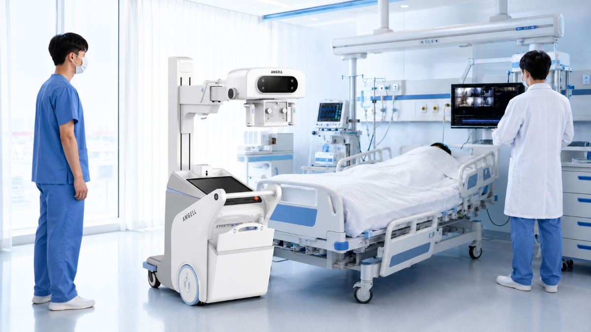
In ICU, emergency, pediatric/NICU, and selected oncology workflows, transport can matter more than fixed-room performance. The strongest use case appears when moving the patient is unsafe or inefficient, so bedside imaging becomes part of the clinical decision rather than a convenience.
Within this workflow, the Lingxi Series supports dynamic observation and high-resolution imaging, including PICC and endotracheal tube placement and verification. Its bedside value also comes from maneuverability and system design: one-handed steering, support for inclines of up to 13 degrees, and a compact dynamic imaging chain built with a wireless dynamic flat-panel detector, compact high-voltage generator, high-heat-capacity X-ray tube, dynamic imaging software, and mobile chassis.
In line and tube workflows, the distinction is practical. A static bedside image can confirm final position; dynamic visualization may also show the route and support adjustment within an approved workflow. Our clinical application materials discuss emergency observation, ICU respiratory motion, pediatric/NICU imaging, oncology line placement, and patients who are difficult to transport; these scenarios show where bedside dynamic imaging can add value, but the final use must match the selected configuration and hospital workflow.
All bedside applications must follow registered indications, local radiation-protection requirements, staff qualifications, and hospital protocols. Mobile access brings imaging closer to the patient; it does not remove clinical or regulatory controls.
Match the Equipment Geometry to the Workflow
Dynamic DR is not one fixed hardware layout. After the clinical need is clear, the next decision is geometry: fixed room, mobile bedside, table-based fluoroscopy, UC-arm positioning, or full-length imaging.
| Main workflow | System direction | Example in our portfolio |
|---|---|---|
| Standardized general radiography | Floor-mounted static DR | MTP Series |
| Dynamic or functional imaging at the bedside | Mobile dynamic DR | Lingxi Series |
| High-volume fixed room with static and dynamic work | Ceiling-mounted dynamic DR | QOMO Series |
| Frequent angled or specialized projections | UC-arm dynamic DR | DTP580 Series |
| GI fluoroscopy and routine radiography on one table | Digital R/F table | Talent II Series |
| Full-spine or full-lower-limb single exposure | Large-format ceiling-mounted DR | Changfeng Series |
These categories and products are part of our current digital radiography portfolio.
Some hospitals are better served by a dedicated static room plus a separate specialized system than by asking one platform to cover every task. Utilization matters more than theoretical feature count.
QOMO combines ceiling-mounted movement, a 4D 10-axis synchronized motion system, and dynamic and static flat-panel detectors. It is intended for fixed rooms that need both routine radiography and dynamic workflows.
DTP580 keeps the tube and detector aligned on a rotating UC-arm, allowing the equipment to change projection while the patient moves less. This geometry suits departments with a varied case mix and frequent angled or specialized views.
Talent II is the more logical route when GI contrast work and table-based fluoroscopy dominate. Changfeng addresses another priority with a proprietary 47-inch detector for full-spine or full-lower-limb imaging in one exposure.
These systems are not interchangeable. The choice depends on whether the main constraint is routine throughput, bedside access, positional range, fluoroscopy workflow, or long-length imaging.
Dose, Training, and Quality Control Belong in the Buying Decision
Dynamic DR should not be described as automatically lower dose than static DR. Dose depends on frame rate, acquisition duration, tube settings, collimation, patient size, repeat rate, and operator technique. Dynamic observation may help avoid some positioning-related repeats, but the sequence itself must be managed by protocol.
Quality control also changes. A respiratory sequence must include the required breathing cycle while the torso remains stable enough for analysis. During a joint or spine motion study, the target anatomy should stay within the field of view and move smoothly through the planned range. Bedside imaging adds shielding, positioning, infection-control, and coordination requirements.
Before purchase, confirm who will create the protocols, how operators will be trained, how dose will be recorded, and how sequences and spot images will move through PACS. Pediatric options, playback tools, reporting, software upgrades, and post-installation application support should be reviewed as part of the workflow package, not only as detector and generator specifications.
What to Send Us Before Requesting a Configuration
A useful review starts with real workflow information. Send us:
- your main examinations and expected daily volume;
- the departments and patient groups the system will serve;
- room dimensions, ceiling conditions, or the bedside transport route;
- the required static, fluoroscopic, dynamic, and functional applications;
- detector, pediatric, DICOM/PACS, dose-recording, training, and local registration requirements.
With this information, our technical and application teams can narrow the choice, identify the required modules, and avoid a quote that includes unused functions or misses an essential workflow. During configuration review, it is useful to provide the case mix, room dimensions, and destination market so the system, documentation, and commercial proposal can be matched to the project.
FAQ
Can a dynamic DR system also take static X-rays?
Many dynamic DR systems support conventional radiography, including systems in our portfolio. The detector arrangement, acquisition modes, and applications still need to be confirmed for the selected configuration.
Is dynamic DR the same as fluoroscopy?
No. Dynamic radiography records a sequence of X-ray images. Fluoroscopy is designed for live or near-live viewing, often during contrast examinations or procedures. Some systems support both, while functional software adds another layer of analysis.
Does dynamic DR always reduce radiation dose?
No. Dose must be assessed using the actual protocol. Frame rate, exposure settings, sequence length, collimation, patient size, and repeat rate all influence the result.
Is functional imaging included with every dynamic DR system?
No. It requires compatible hardware, acquisition protocols, software modules, and approved clinical indications. These items should be listed explicitly in the proposed configuration.
When is mobile dynamic DR preferable to a fixed system?
It is especially relevant when patient transport, bedside positioning, line or tube observation, or respiratory motion assessment is part of the clinical problem. A fixed room cannot bring imaging to a patient who should not be moved.
Conclusion
In the end, the choice is straightforward: use static DR when a well-positioned still image answers the question; choose dynamic DR when the examination depends on motion, live confirmation, bedside access, contrast flow, or functional analysis. A good configuration should fit the case mix, room setup, operators, and integration plan instead of chasing the longest specification list.