
Introduction
Both C-arm and U-arm systems use X-rays. Both produce diagnostic-quality images. Yet they serve different stages of patient care, occupy different rooms, and move in fundamentally different ways. Choosing the wrong geometry wastes floor space, slows clinical workflow, and forces staff to work around the equipment rather than with it.
We break down the structural, clinical, and operational differences between a mobile C-arm and a U-arm (UC-arm) system, using specific product examples to keep the comparison grounded in equipment that actually exists on the market.
C-Arm vs U-Arm at a Glance
| Factor | C-Arm X-Ray System | U-Arm / UC-Arm DR System |
|---|---|---|
| Primary role | Imaging during a procedure | Imaging for diagnostic examinations |
| Typical location | Operating room, procedure room, urology suite | Radiography room, examination centre, occupational-health setting |
| Mobility | Wheeled to the procedure table | Installed at a dedicated imaging station |
| Main imaging mode | Live fluoroscopy and frame capture | Multi-position radiography; fluoroscopy on dynamic models |
| Patient positioning | Equipment approaches the patient and table | Arm and detector move to reduce patient repositioning |
| Common workload | Fixation checks, guidewire/catheter navigation, stone and stent procedures | General examinations, specialised projections, stitching, contrast studies, pneumoconiosis screening |
| Room planning | Table clearance, movement route, storage, detector access | Installation footprint, rotation envelope, patient access, workstation placement |
The practical rule: choose a C-arm when imaging must follow an active procedure at the table. Choose a U-arm or UC-arm when one diagnostic room must cover varied projections and examination types.
What Is a C-Arm X-Ray System?
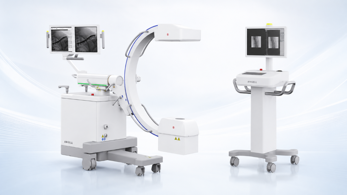
A mobile C-arm is a wheeled X-ray unit whose source and detector sit at opposite ends of a C-shaped arm. The assembly can be positioned around the patient while the operating table and sterile field stay in place — giving the surgical team live fluoroscopic feedback as the procedure advances.
That live feedback matters because the image changes as the procedure progresses. During fracture reduction, the team may check alignment and screw direction at several stages. Vascular work requires guidewire and catheter navigation under continuous imaging. Urology workflows span lithotripsy, ureteroscopic stone removal, percutaneous access, and stent placement. Emergency and trauma cases add a speed requirement: the system must enter the room, reach the table, and begin imaging within a tight setup window.
A C-arm can also capture still frames when a clinically important position needs to be documented, combining live observation with position confirmation during treatment.
Flat-Panel Detector vs Image Intensifier — Why It Matters
Not every C-arm is built the same way. The detector technology sitting at the receiving end of the arm has a direct impact on image geometry, working space, and long-term cost of ownership.
| Flat-Panel Detector (FPD) C-Arm | Image Intensifier (I.I.) C-Arm | |
|---|---|---|
| Image field shape | Rectangular — the full sensor area is usable | Circular — display corners carry no image |
| Geometric distortion | Flat sensor: no pincushion or S-distortion | Curved input surface introduces pincushion and orientation-dependent distortion |
| Brightness uniformity | Uniform response across the field | Vignetting — brightness falls off toward the edges |
| Performance over time | No intensifier tube to age; output stays stable | Intensifier gain degrades, typically pushing dose up to maintain image quality |
| Form factor | Slim detector housing leaves more room at the table | Deep, bulky intensifier housing on the receiving end |
When evaluating a C-arm, this is the first technical fork in the road. FPD C-arms may carry a higher upfront purchase price than image-intensifier systems, but they avoid the detector aging and image-degradation issues associated with intensifier technology.
What Is a U-Arm or UC-Arm X-Ray System?
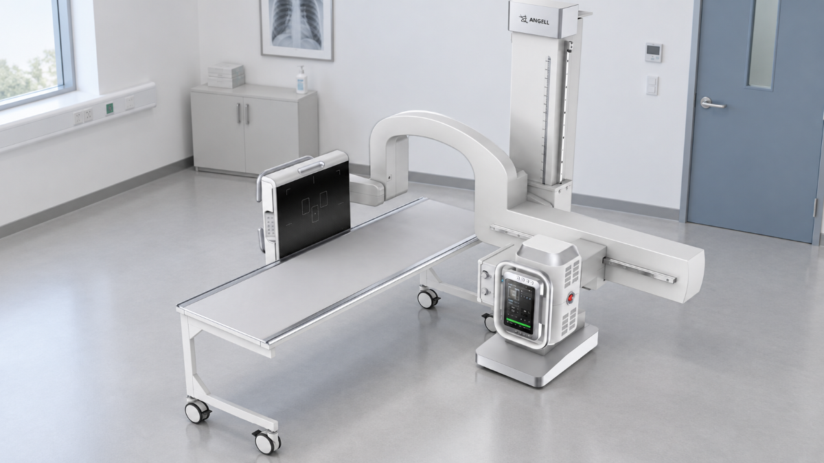
A UC-arm dynamic DR system places the X-ray tube and flat-panel detector on one connected mechanical structure. The name describes the geometry: at rest the arm forms a U; once rotated into a working projection, it resembles a C. Because tube and detector ride the same structure, source-to-image distance and beam centring arrive already correct with every position the operator selects.
Changing the projection means rotating the arm — not moving the patient. That distinction matters in rooms where the next case is rarely like the last one. The system can support upright, supine, oblique, axial, and cross-table lateral studies while setup adjustments happen on the equipment side rather than the patient side.
Capability varies by model. A static U-arm captures still radiographs only. A dynamic UC-arm adds fluoroscopy, contrast-enhanced imaging, stitched full-body views, and rapid dynamic/static switching — turning one room into a multi-function diagnostic station.
Five Key Differences Between C-Arm and U-Arm
1. The Clinical Decision the Image Supports
A C-arm serves decisions made during treatment: whether a reduction is acceptable, whether an instrument has reached the target, whether a guidewire is tracking as planned. A UC-arm serves a broader diagnostic question across body regions and projections. Dynamic imaging may be available on both, but the clinical reason for using it is different.
2. Where the Equipment Lives
A mobile C-arm travels to operating and procedure rooms; movement routes, storage position, and table access all factor into the room plan. A UC-arm is installed in a dedicated radiography room and stays there — supporting a wider examination range from one fixed station.
3. How Projection Changes Happen
With a C-arm, the assembly moves around the procedure table while clinicians preserve access to the sterile field. With a UC-arm, the connected source-detector pair rotates or travels along the arm to create the next projection. That mechanical difference affects table compatibility, collision clearance, patient transfer, and where the operator stands.
4. Departments and Examination Mix
The department name identifies the service line; the imaging task determines the system. Orthopaedics may need a mobile C-arm for fixation at the operating table and a UC-arm for standing or recumbent diagnostic imaging. Emergency services may use a UC-arm for diagnostic reception and a mobile C-arm during trauma surgery. The two systems address different moments of the same patient journey.
5. Image Acquisition and Workflow
In a C-arm workflow, fluoroscopy provides immediate feedback at the table. Operators position the system, monitor the live image, and capture key frames when instrument or implant position needs to be recorded.
In a dynamic UC-arm workflow, the focus shifts to switching between examination types — radiography, fluoroscopy, contrast studies, stitched imaging — reviewing results at the workstation, and routing them to PACS. During equipment evaluation, hospitals should test live fluoroscopy and table-side capture for a C-arm, and mode switching, stitching quality, and PACS integration for a UC-arm.
Matching the System to the Clinical Scenario
These differences become easier to apply when they are tied to a specific clinical setting. A department that performs both image-guided procedures and diagnostic examinations should plan the two types of work separately. The following scenarios show how the choice changes with the clinical task, patient position, and the room in which imaging takes place.
Orthopaedic Surgery vs Orthopaedic Diagnosis
Fracture reduction, fixation, and implant checks require live imaging that follows the operation — a mobile C-arm. Standing or recumbent diagnostic studies, including full-spine or full-lower-limb stitching and specialised joint projections, belong in a UC-arm room. A department performing both should budget for both.
Emergency Reception vs Emergency Surgery
Diagnostic X-rays completed in the imaging room fit a UC-arm workflow, especially when the system must handle standing, recumbent, or limited-mobility positions without patient repositioning. Once the patient enters trauma surgery and the team needs live guidance, the workflow shifts to a mobile C-arm.
Urology and Vascular Procedures
In urology and vascular procedures, the deciding factor is whether clinicians need to watch a device or contrast medium move during the procedure. Guidewire, catheter, stent, instrument, or contrast tracking calls for a mobile C-arm. After that category is selected, the configuration review should focus on detector field of view, arm depth, table clearance, and dose-management functions.
Physical Examination and Occupational Health
Physical examination and occupational-health rooms often handle varied body regions and projections throughout the day. A UC-arm configuration should be checked against the required full-spine or full-lower-limb imaging, Waters, patella axial, calcaneal axial, cross-table lateral, and any fluoroscopy or stitching needs. For pneumoconiosis programs, our DTP580 adds dedicated image processing for that workflow.
How These Differences Show Up in Real Equipment
These application differences can be seen in our HUA II and DTP580. The examples below show how each system supports the procedural or diagnostic use described above.
HUA II Series — Mobile Dynamic FPD C-Arm
The HUA II was designed around the idea that no two procedure rooms are set up the same way. It converts between all-in-one and split configurations — an industry first for a dynamic FPD C-arm — so the system layout can match the table position, room shape, and operator preference without swapping equipment.
Six low-dose safety features work together during every procedure: Auto Exposure, Automatic Programme Response (APR), laser positioning, Automatic Brightness Stabilisation with Instant Dose Control (ABS + IDC), Dose Area Product display and analytics, and remote exposure. Rather than treating dose management as a single toggle, the system layers multiple controls across the imaging chain.
The dynamic flat-panel detector supports implant-position adjustment under fluoroscopy and post-operative assessment, with all-format image export and multi-frame acquisition modes.
DTP580 Series — Multifunctional UC-Arm Dynamic DR
The DTP580 addresses a different problem: a single diagnostic room that must cover a broad, unpredictable case mix. Both the detector and the horizontal arm rotate independently, enabling Waters view, patella axial, calcaneal axial, and cross-table lateral projections to be set up on the equipment rather than negotiated with the patient.
The system integrates radiography, fluoroscopy, contrast-enhanced imaging, full-body stitching, and pneumoconiosis examination on one platform. Rapid dynamic/static switching, video playback, and PACS integration keep the workflow moving between examination types without leaving the room.
For occupational-health programmes, self-developed DR image-processing software provides standardised pneumoconiosis examination output — a capability built specifically for that clinical protocol rather than adapted from general-purpose tools.
What Sits Behind Both Systems
Both the HUA II and the DTP580 draw on the same vertically integrated engineering base. Our Technology develops its own flat-panel detectors, X-ray tubes, and high-frequency high-voltage generators — giving the design team control over the full imaging chain rather than assembling third-party components.
That integration started with the company's founding in 2002 and installed base has reached 30,000 units across more than 100 countries. In 2014, we introduced the world's first UC-arm dynamic DR system equipped with a flat-panel detector — a milestone that marked the transition from image-intensifier-based U-arm designs to the FPD-based architecture used in the DTP580 today.
Selection Checklist
Use this as a starting framework. The final specification depends on the exact system model, room conditions, and local regulatory requirements.
| Decision Point | Points Toward a Mobile C-Arm | Points Toward a UC-Arm |
|---|---|---|
| When does imaging happen? | During surgery or a procedure | Before or after — diagnostic workup, follow-up, screening |
| Does the image need to be live? | Yes — fluoroscopy guides the procedure | Sometimes — dynamic models add fluoroscopy, but the primary workload is radiography |
| Where is the patient? | On the operating or procedure table | Standing, seated, supine, or limited-mobility in the imaging room |
| How often does the projection change? | Repositioning happens around the sterile field | Frequently — different body region or angle for the next patient |
| What must the room accommodate? | Entry route, table clearance, storage, staff access | Installation footprint, rotation envelope, workstation, PACS connection |
| Is there a specialised screening programme? | Not typically | Pneumoconiosis, full-body stitching, occupational-health protocols |
Common misconception worth noting: a UC-arm that includes fluoroscopy does not replace a surgical C-arm. Surgical use also requires table-side mobility, sterile-field access, appropriate arm depth, and repeated repositioning around the patient during treatment. Likewise, a mobile C-arm in any room still requires planning for entry routes, storage, table clearance, monitor placement, and the system's full range of movement.
FAQs
Is a U-arm the same as a C-arm?
No. At certain angles the shapes can look similar, but the installation, movement envelope, and core clinical workflow are fundamentally different. A C-arm is mobile and follows procedures. A U-arm is installed and supports diagnostic examinations.
Can a department need both?
Yes. A hospital may use a mobile C-arm for intraoperative guidance and a UC-arm for multi-position diagnostic imaging. The systems serve different stages of care, so fluoroscopy capability alone does not make them interchangeable.
What information is needed before requesting a configuration?
For a mobile C-arm: planned procedures, operating-table layout, available working clearance, detector field-of-view requirements, and dose-management priorities. For a UC-arm: examination mix, room dimensions, required projections, stitching and occupational-health needs, PACS environment, and installation country.
Does a U-arm require more room than a C-arm?
There is no universal answer. A UC-arm needs an installation footprint and rotation envelope. A mobile C-arm needs travel routes, storage space, and working clearance around the table. The room plans are different in kind, not simply in size.
Conclusion
The right system depends on the clinical task, the room it occupies, how patients are positioned, and the imaging modes the department actually uses. A mobile C-arm supports procedural imaging at the table. A UC-arm supports multi-position diagnostic work in the radiography room. Hospitals that perform both should plan each workload on its own terms.
Share your procedure list or room plan with us, and the technical team will review the configuration against the intended clinical use.