
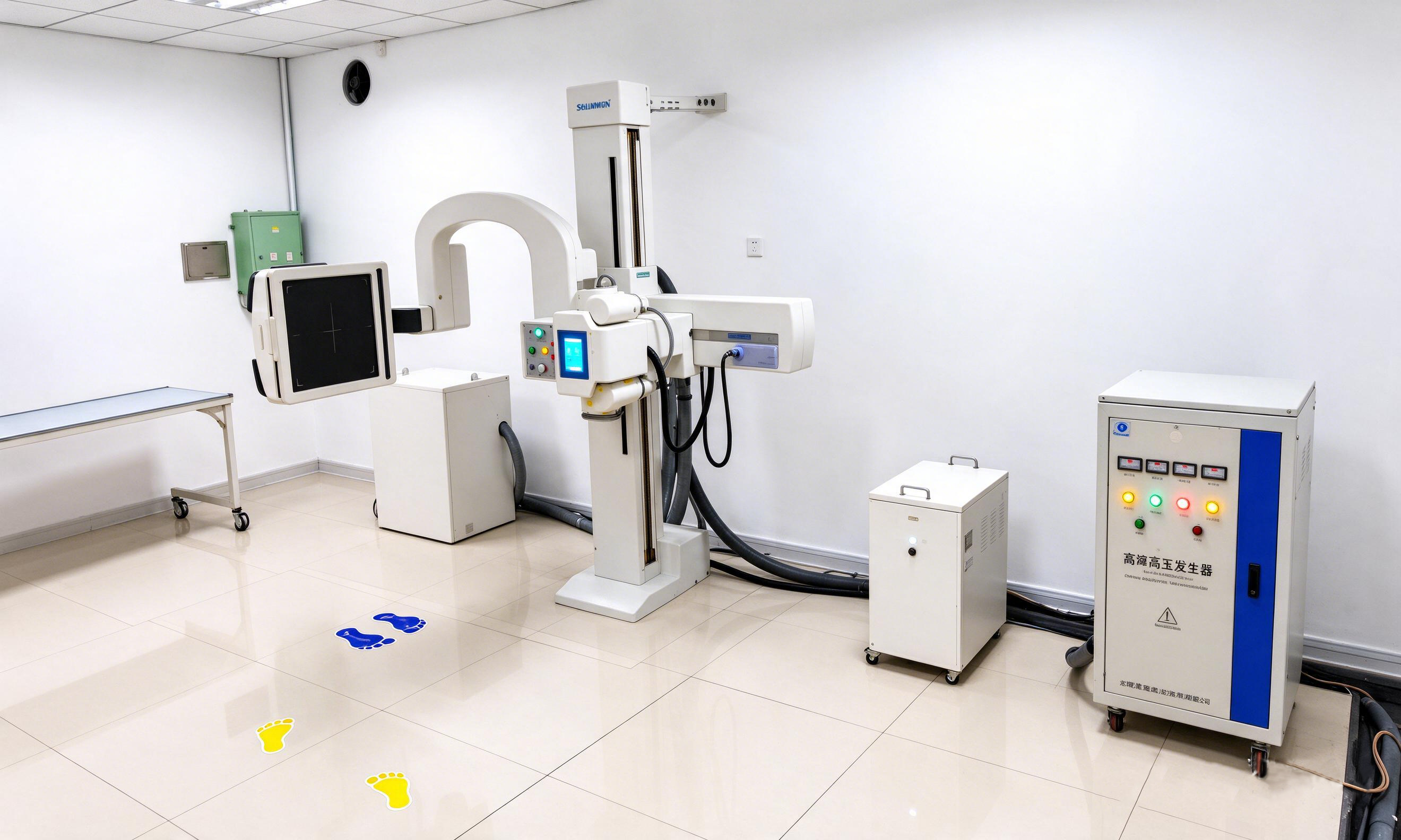
Lumbar spine X-rays are used to evaluate lower back symptoms, suspected injuries, and changes over time. A routine examination typically includes AP and lateral views, with oblique views added for specific clinical indications. Patient positioning, receptor placement, and X-ray beam alignment vary by projection to capture the required anatomy.
Confirm the Requested Views Before Positioning
Before positioning, the technologist needs a clear view of the required anatomical coverage, projection, and any patient limitations.
Projection | Positioning goal |
|---|---|
AP | Requested lumbar anatomy centered with minimal rotation. |
Lateral | True lateral alignment maintained from the shoulders through the pelvis. |
Oblique | Correct side and rotation, with an accurate anatomical side marker and a projection label matching the patient’s position. |
AP, Lateral, and Oblique Lumbar Spine Positioning
Routine lumbar spine views are generally taken at the end of expiration, with the patient holding their breath during exposure.
AP Lumbar Spine Positioning: Set the Requested Coverage
1. Fit the receptor to the prescribed anatomy.
The image receptor must cover all the required anatomy, which can extend from T12 to the lower sacrum, including the sacroiliac joints, as outlined in the Dartmouth Hitchcock protocol.
A larger lumbar or lumbosacral field typically uses a 35 × 43 cm receptor centered at the iliac crest. Smaller lumbar fields may be centered closer to L3. The iliac crest is approximately level with L4 or L4–L5.
If more of the sacrum needs to be included, the receptor may need to sit lower while still covering the upper lumbar spine. Widening the collimation alone will not capture anatomy outside the detector.
2. Align the patient and beam with that field.
For a routine AP view, the patient lies on their back with the spine centered over the receptor. Slight flexion of the hips and knees helps reduce lumbar lordosis (the natural inward curve of the lower back) and improves comfort.
Patients with suspected spinal injuries require extra care to maintain spinal alignment and minimize movement. Their legs should remain in a stable, supported position that follows spinal precautions.
The shoulders and pelvis should be level to minimize rotation. The central X-ray beam is perpendicular to the receptor and centered on the required lumbar anatomy.
Lateral Lumbar Spine Positioning: Establish a True Lateral Position
For a routine lateral view, the patient lies on either side with the shoulders and hips stacked. The waist is supported with a radiolucent pad to keep the lumbar spine parallel to the receptor. The pad should fill the gap between the waist and the table without raising the spine too high.
Slightly bent hips and knees help the patient stay comfortable. Padding between the knees reduces pelvic rotation, and the arms rest in front of the body, outside the imaging field.
The image receptor is positioned lengthwise alongside the lumbar spine, centered near L3. The central X-ray beam is perpendicular to the receptor, approximately 4 cm above the iliac crest and 5 cm forward from the posterior skin surface. A compensating beam angle can help correct any remaining lumbar tilt, but shoulder or pelvic rotation requires an adjustment to the patient's position.
Supine cross-table lateral positioning for patients with injuries
Patients with suspected spinal injuries may need to remain on their backs to avoid unnecessary spinal movement. A supine cross-table lateral view allows imaging without turning the patient onto their side.
The shoulders and hips remain aligned, with the receptor positioned vertically beside the lumbar spine. The X-ray beam travels horizontally through the patient toward the receptor, centered on the required lumbar anatomy. The arms can rest across the upper chest if this can be done safely.
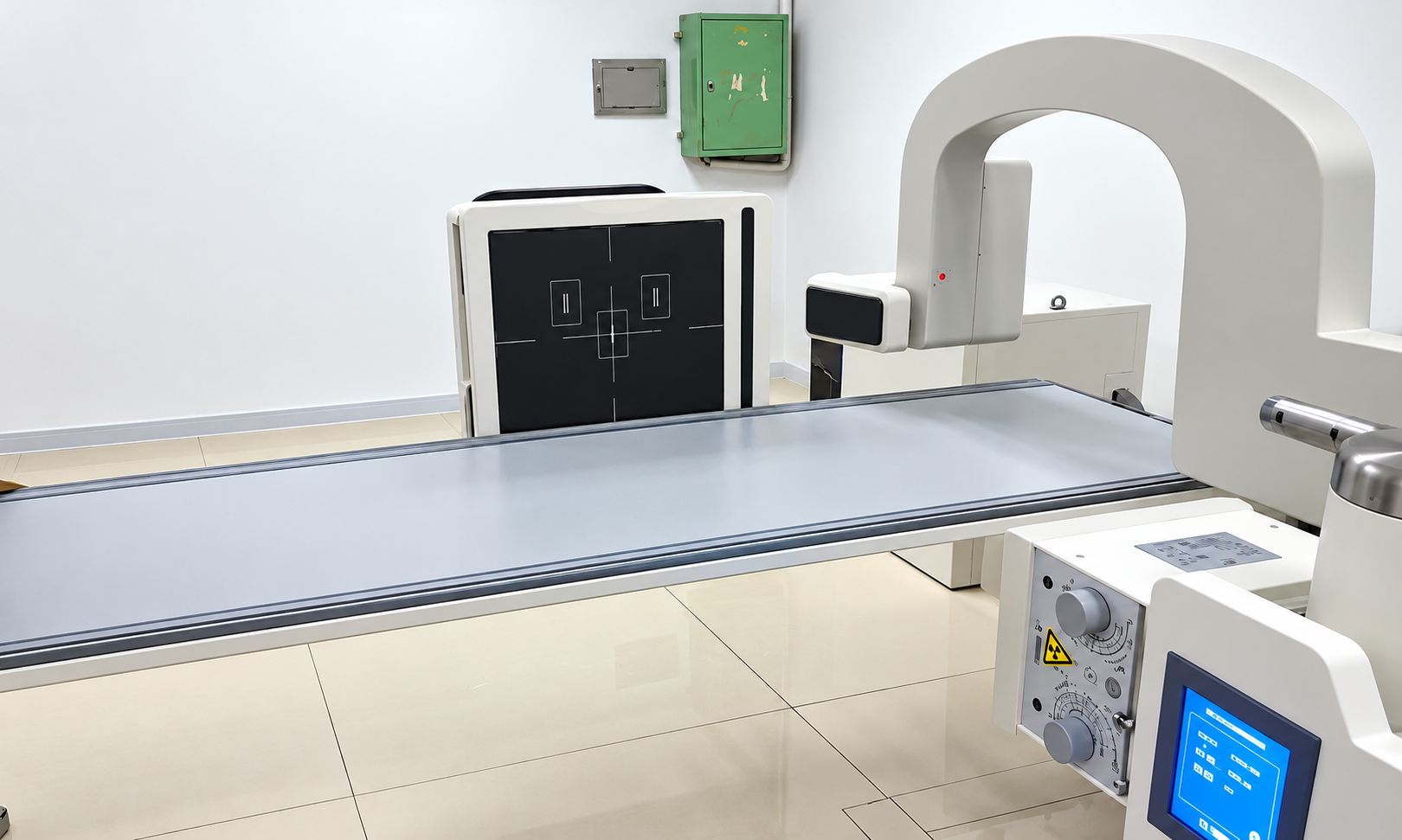
For this type of imaging, DTP580 allows the detector and horizontal arm to rotate into position for a cross-table lateral projection.
Oblique Lumbar Spine Positioning: Control the Selected Rotation
For posterior oblique views, the patient's position determines which zygapophyseal (facet) joints are shown:
Requested projection | Patient position | Joints demonstrated |
|---|---|---|
RPO | Right posterior side nearer the receptor; left side elevated | Right zygapophyseal joints |
LPO | Left posterior side nearer the receptor; right side elevated | Left zygapophyseal joints |
The anatomical side marker identifies the patient's right or left side, while RPO and LPO indicate the patient's position. The projection label must match the actual position. Bilateral posterior oblique views require both RPO and LPO positions. PA oblique views use a different positioning method.
For a routine posterior oblique view, the patient lies on their back and rotates the trunk and pelvis together to approximately 45°. A support under the raised side helps maintain the position. The rotation angle depends on the lumbar level and patient anatomy.
The central X-ray beam enters approximately 5 cm medial to the elevated anterior superior iliac spine (ASIS) and 4 cm above the iliac crest, near L3. The beam is perpendicular to the receptor, which is centered beneath the lumbar region.
Spinal injuries, severe pain, or limited mobility can make rotation difficult or unsafe. These patients should not be forced into an oblique position. The oblique view may need to be modified or omitted based on the patient's condition and examination requirements.
Review the image before repeating an exposure
The need for a repeat X-ray depends on whether the required anatomy is clearly visible. Each projection has different signs of correct positioning.
AP view: The lumbar spine should appear near the image center, with spinous processes aligned and pedicles reasonably symmetrical. Missing anatomy or marked rotation can affect interpretation.
Lateral view: Closely overlapping posterior vertebral body margins indicate minimal rotation. The iliac crests provide another reference for checking alignment. Noticeable separation suggests the patient was not positioned in a true lateral view.
Oblique view: The main concern is whether the intended facet joints and pars interarticularis are clearly visible. Poor visualization may indicate incorrect rotation, affecting the image's suitability for diagnosis.
Frequently Asked Questions
Can one lumbar spine view replace another?
No. AP, lateral, and oblique projections serve separate positioning purposes. Each uses a different patient and receptor relationship and addresses a different positioning question. After an AP image is acquired, any separately requested lateral or oblique view remains part of the examination.
What should be done when the examination order and local protocol do not match?
When the examination order and local protocol specify different view sets, the technologist checks the discrepancy with the radiologist or designated protocol lead and confirms which views to acquire, modify, or omit before proceeding.
What should be documented when a standard positioning method is modified?
Changes made because of trauma, pain, limited mobility, or patient safety belong in the examination record. The entry names the projection performed and explains why the routine method changed. This gives the interpreting team context when the image differs from the routine setup.
Conclusion
A lumbar spine X-ray is planned from the examination request. The anatomy range, patient condition, projection, and local protocol determine the setup, and any change in those factors calls for a positioning reassessment before exposure. Where a general positioning reference differs from the department’s approved protocol, institutional protocol and the patient’s condition take priority.
Our UC-ARM Dynamic DR workflow shows how detector movement supports different projections while reducing the need to reposition patients across a range of examinations. At Angell, our team can then review the workflow requirements with you.