
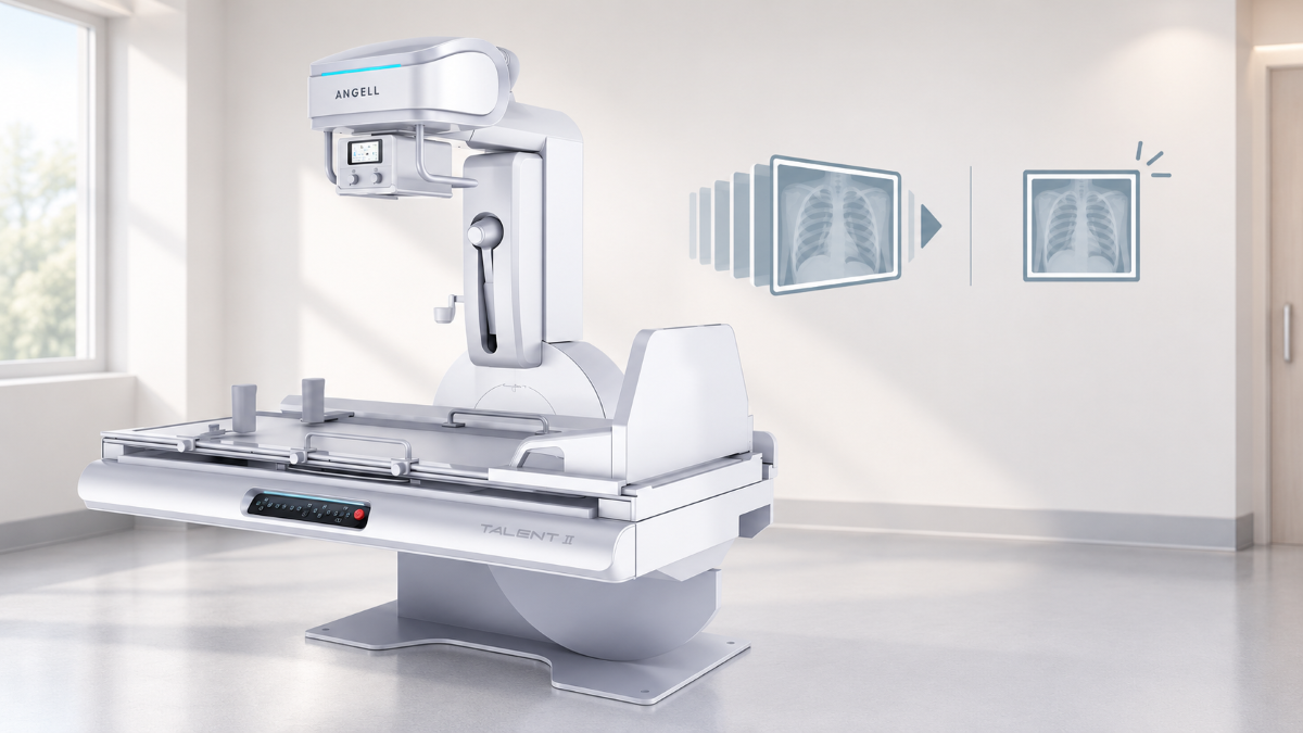
The core difference in fluoroscopy vs radiography is the output. Radiography records static X-ray images, while fluoroscopy shows a live, continuous or rapidly pulsed X-ray sequence so clinicians can watch movement, contrast flow or a device as an examination unfolds. A fracture, a lung field or an implant check can be assessed from a still picture; a swallow, a catheter route or the steps of a fracture reduction usually require imaging as the action happens.
Three common misconceptions blur that line: that fluoroscopy always requires contrast, that it always uses a continuous beam, and that the two modes require separate rooms. In practice, fluoroscopy can be performed without contrast, many systems use pulsed acquisition, and R/F or suitably configured dynamic DR platforms can deliver both modes in one room.
Fluoroscopy vs Radiography at a Glance
Comparison point | Radiography | Fluoroscopy |
|---|---|---|
Primary output | A static projection or a set of projections | A real-time or near-real-time image sequence |
Question it answers | What does the anatomy look like at a chosen moment? | What happens as anatomy, contrast or a device moves? |
Typical examinations | Chest, bones, joints, abdomen, alignment and follow-up | Swallowing studies, GI contrast work, device guidance and selected procedures |
Exposure pattern | Separate, controlled exposures | Continuous or rapidly pulsed acquisition |
Contrast | Used in selected examinations | Common in some studies, not part of the definition |
What gets saved | Static images | Sequences, selected frames and spot images |
Main dose drivers | Projection, technique factors and number of views | Beam-on time, pulse rate, field size and saved acquisitions |
Equipment | Radiography rooms and mobile DR | Fluoroscopy rooms, R/F tables, dynamic DR systems and C-arms |
US federal regulations draw the same line. Under 21 CFR 1020.30, radiography records an X-ray pattern so the user gets an image after the exposure ends, whereas fluoroscopy presents images continuously as they are generated.
How Each Mode Produces Its Images
Radiography: One Exposure, One Image
A radiograph comes from a single, short exposure. The radiographer positions the patient for a defined projection, sets the technique or lets automatic exposure control end the exposure, and the detector records the attenuation pattern once. Because the beam is on for only a fraction of a second, the exposure can be set for fine detail and low noise, and motion blur stays limited. The result is a still image that can be measured, compared with earlier studies and archived. A multi-view chest examination repeats the static acquisition for each projection. Long-length radiography may instead use multiple stitched exposures, slit scanning or a single large-format exposure, depending on the system.
Fluoroscopy: Repeated Frames While the Beam Is On
Fluoroscopy produces a stream of images while the beam is on, typically under foot-switch control, using either continuous output or short pulses. In pulsed mode, the detector reads each frame fast enough for the monitor to show smooth motion. Fluoroscopic frames are typically acquired at a lower dose per frame than a diagnostic radiograph. Image noise becomes more visible when less radiation reaches the detector, although detector performance and image processing also affect the result. Automatic brightness control adjusts the technique as the patient or beam angle changes. When the operator releases the switch, last-image hold keeps the final frame on screen, so positioning can be checked without further exposure.
Spot Images, Saved Sequences and Radiographs
A fluoroscopic examination may save a moving sequence, a last-image-hold image and selected spot or acquisition images. How those still images are produced varies by system and acquisition mode. A saved sequence stores the moving frames for replay, which matters when the motion itself is the finding, such as contrast entering the airway during a swallow. A last-image-hold image is the final live frame kept after the beam stops, and US regulations allow it to be either a retained fluoroscopic frame or a separate radiographic exposure triggered as fluoroscopy ends. A spot image is a still captured at a chosen moment during the study, and many systems acquire it as a short radiographic exposure so it carries more detail than a single live frame. In a barium swallow, for instance, the saved sequence can show aspiration as it happens, and spot images document the shape of a narrowing.
A conventional radiograph differs from all three because the projection is planned in advance. Reproducing the same projection and patient position at follow-up helps clinicians compare changes over time.
Which Clinical Questions Each Mode Answers
Radiography Answers Questions About Structure
Radiography fits any decision that can be made from a still view of the anatomy. Is the wrist fractured? Is the knee aligned? Does the lung field show consolidation? Has a screw stayed where it was placed, and has anything changed since the last study? Each answer comes from structure at a defined moment.
A radiographic study may include several projections or a stitched long-length spine image, but each exposure is still reviewed as a static picture. That makes radiography well suited to follow-up and alignment comparisons.
Fluoroscopy Answers Questions About Movement and Route
Fluoroscopy is the appropriate mode when the finding depends on a path, timing or changing position. A swallowing study shows how a bolus moves and whether any of it enters the airway. A GI contrast examination shows filling, emptying and peristalsis. In the operating room, live imaging lets a surgeon watch a fracture reduce and adjust an implant before closing, and catheters, guidewires and needles are followed as they advance during angiographic and urological work.
Some of this work involves no contrast at all. Watching diaphragm motion or checking a tube during placement relies on the moving image alone.
When One Examination Uses Both Modes
Several diagnostic studies move between the two modes within minutes. During a barium swallow, the radiologist watches contrast in real time and captures spot images at the moments worth retaining. A hysterosalpingogram follows contrast through the uterus and fallopian tubes under fluoroscopy, with still images recorded once filling is visible. The same pairing can extend across a patient pathway: fluoroscopy guides a fracture reduction in theatre, while radiographs document healing during follow-up.
Where studies like these make up a regular share of the workload, an R/F system or suitably configured dynamic DR system can support both modes in one room. Talent II illustrates that arrangement: its 17 x 17-inch dynamic flat-panel detector supports radiography and fluoroscopy on the same R/F table, alongside GI contrast studies and long-length stitching. If live observation is rare and most examinations are chest, spine or extremity studies, a radiography-only room avoids the protocols, training and cost of an underused fluoroscopy capability.
What the Patient Experiences
The two modes also feel different to the patient. For a radiograph, the patient is positioned, asked to keep still and perhaps hold a breath, and the exposure is over almost instantly. Many diagnostic fluoroscopy examinations involve additional preparation or participation. Depending on the study, the patient may drink barium or receive contrast through a catheter or injection, turn or change position on a tilting table, and swallow or breathe on instruction while the radiologist watches the screen. These examinations generally take longer than routine radiographs, and an upper GI series may require fasting beforehand. Local staffing practices vary, but diagnostic fluoroscopy often involves both a radiologist and a radiographer at the table, whereas routine radiographs are commonly acquired by a radiographer.
Radiation Dose: Fluoroscopy Is Usually Higher Than a Single Radiograph
A fluoroscopic examination tends to deliver more radiation than a single radiograph because it produces many frames over time. Approximate adult reference values in the Merck Manual put a two-view chest X-ray at about 0.1 mSv, a single abdominal X-ray at 0.7 mSv and an upper GI study with barium at about 6 mSv. The same table lists 1.5 mSv for one lateral lumbar-spine view. These are different examinations, and the table notes that doses vary, so the figures show a range of typical values and cannot predict the dose of a specific protocol.
A fair comparison holds the procedure and the patient group constant. Within fluoroscopy, beam-on time, pulse rate, field size, magnification, patient size, procedure complexity and the number of recorded images all affect the final dose. In a 2011 bench simulation published in the Korean Journal of Pain, researchers used a lead apron in place of a patient and a 15-fps pulsed setting. Pulsed mode reduced the radiation-absorbed dose by 32%, low-dose mode by 57%, and the two together by 83% relative to conventional mode. Those percentages come from one test setup and do not transfer automatically to every platform, patient or protocol.
The two modes are also tracked differently. A radiograph is described by its technique factors and, on many systems, a dose-area product (DAP) value for each exposure. For fluoroscopic systems manufactured on or after June 10, 2006, US federal performance standards require displays of air kerma rate and cumulative air kerma. Departments may also record fluoroscopy time and DAP under their own protocols. These values support comparisons between procedures performed under similar conditions.
The FDA notes that doses can run relatively high, especially in complex interventional procedures with long beam-on times where skin injury becomes a concern, and that the probability of such effects is statistically very small. Contrast use does not determine radiation dose by itself, and pulsed acquisition can lower exposure without eliminating it.
Dose does not usually determine the imaging mode; the clinical question does. When one or more static views can answer it, radiography is generally the lower-dose route. When the study relies on motion, contrast tracking or device guidance, fluoroscopy is justified, and the work shifts to keeping the protocol lean: lower pulse rates where image quality allows, tight collimation, last-image hold in place of repeated screening, and a recorded DAP or air kerma value for each study. If a static study cannot answer the question, additional or alternative imaging may still be needed.
When fluoroscopy is used to guide a device or implant, exposure control and dose information need to be available during the procedure. The HUA II Series is a mobile dynamic-FPD C-arm that combines automatic brightness stabilization and instant dose control with DAP display and analytics. These functions support real-time adjustment and dose review, while beam-on time, pulse rate and collimation still depend on the protocol and operator practice.
Can One System Handle Both Modes?
Yes. The clinical question determines the imaging mode; patient access, room geometry and department workload determine the platform. Radiography and fluoroscopy are imaging modes, while R/F tables, dynamic DR systems and C-arms are platforms that deliver one or both in different settings.
R/F, Dynamic DR and C-Arm Are Not Interchangeable
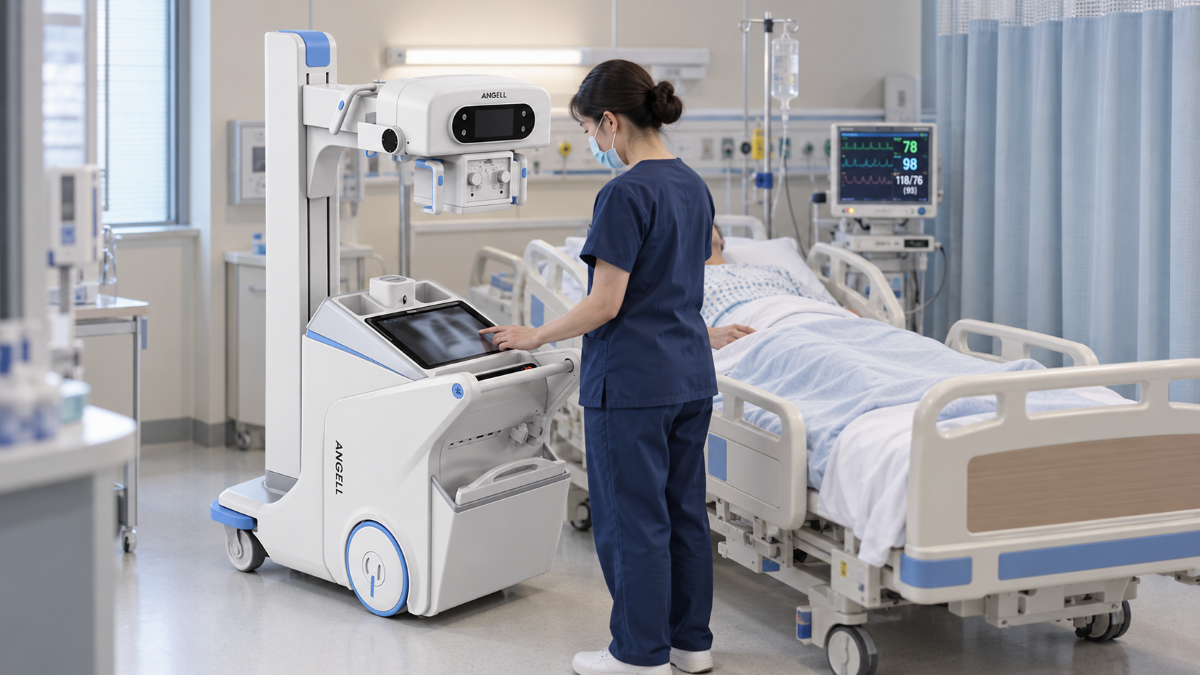
An R/F system combines radiography and fluoroscopy on a table built for diagnostic work, mainly GI, GU and other contrast studies. Dynamic DR describes a DR system that can also acquire image sequences, and whether a given model supports full fluoroscopy, contrast studies or functional analysis varies by configuration. A mobile C-arm is built around procedural access, with an arm that wraps around the patient and operating table. A dynamic DR unit that shows live images at the bedside is therefore not a surgical C-arm, and a mobile C-arm is not designed to replace the positioning range or routine workflow of a general radiography room. Our guides to dynamic DR vs. static DR and C-arm vs. U-arm examine those two boundaries in more detail.
Combining both modes also asks more of the detector. Live frames need fast readout and good sensitivity at a low dose per frame, while a spot image or radiograph needs fine detail from one exposure. Some combined rooms use a single dynamic flat-panel detector for both, so the study runs on one imaging chain from live screening to spot image. Others pair a dynamic panel with a separate static panel and switch between them during the examination. The choice matters most in departments with daily contrast lists and frequent spot imaging, and much less where fluoroscopy is occasional.
Fixed Room, Bedside or Operating Room
Where the patient can be imaged narrows the choice further. A fixed-room R/F table suits departments that run GI and contrast lists alongside routine radiography. It provides table-based positioning, room-side controls and a stable workflow for repeated contrast examinations, but it depends on the patient being brought to the room.
Transferring ventilated ICU patients, premature infants or patients undergoing oncology care may be difficult or clinically undesirable. The Lingxi Series brings dynamic observation, high-resolution spot imaging and sequence playback to the bedside for PICC and endotracheal tube placement checks.
Operating-room procedures introduce sterile-field and access requirements. A mobile C-arm provides live imaging around the operating table without trying to replace the positioning range of a general radiography room. Fixed R/F, mobile dynamic DR and C-arm systems therefore solve three different access problems rather than three versions of the same one.
Frequently Asked Questions
What is the main difference between radiography and fluoroscopy?
Radiography records a static image after the exposure ends. Fluoroscopy displays an image sequence as it is generated, allowing clinicians to follow motion, contrast or a device during an examination.
Does fluoroscopy always require contrast?
No. Contrast is common in GI, urological and vascular studies, and fluoroscopy also runs without it, for example, when guiding fracture reduction or watching diaphragm movement.
Which has the higher radiation dose?
A complete fluoroscopic study usually exceeds a single radiograph. Meaningful comparisons use the same procedure and patient group and look at beam-on time, pulse rate, field size and the recorded dose indicators, not the modality label alone.
Can one X-ray system perform both modes?
Yes. R/F systems and dynamic DR systems configured for fluoroscopy, support both modes on one platform. The exact modes, detector arrangement and software modules are set by the configuration.
Final Decision Rule: Static Imaging, Live Imaging or Both
Use radiography when static projections answer the clinical question and fluoroscopy when the decision depends on motion, contrast flow, or device guidance. If a department regularly needs both, choose the platform only after mapping patient mobility, required positions, room geometry and daily workload to a fixed-room, bedside or operating-room workflow.
Founded in 2002, Angell develops digital medical imaging systems and related imaging-chain technologies, including X-ray detectors, tubes, generators, DR platforms and image acquisition software. Discuss Your Department Workflow With Our Team