Digital Mammography System medical imaging solutions

Digital Mammography System

Product Categories

Product Category

Digital Mammography Systems

Full-field digital mammography for breast cancer screening.

A digital mammography system is a dedicated low-energy X-ray unit built around a single anatomy. Everything in it — the tube spectrum, the compression paddle, the detector pitch — exists to separate soft tissue that differs in density by only a few percent, and to find calcifications measured in fractions of a millimetre.

In full-field digital mammography (FFDM), a flat-panel detector replaces the film cassette. The exposure is no longer the finished image: it is data that can be windowed, magnified, sent to PACS and compared against the same patient's prior years. That single change is what turned breast imaging from a darkroom procedure into breast cancer screening equipment a health system can run at population scale.

In this category

Fanghua Series

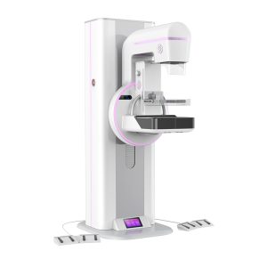
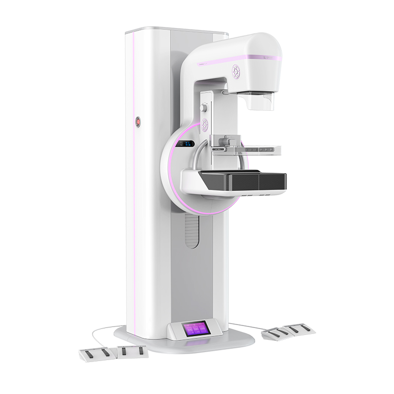
Angell's innovative digital mammography system. A large-format flat-panel detector covers the breast, lymph nodes and axilla in one field; tomosynthesis is standard; and dose is managed by an intelligent motorized collimator together with automatic exposure control. Detector format, tube configuration, tomosynthesis behaviour and dose options are set out on the product page.

Explore Fanghua Series Get a quote

The technology

What full-field digital changed

Screen-film mammography produced excellent images — once. Its limit was never sharpness; it was that acquisition, display and storage were locked into one piece of physical film, so a single exposure had to be right for fat, glandular tissue and calcifications at the same time. FFDM breaks those three jobs apart.

Screen-film mammographyFull-field digital mammography
Image captureFilm is receptor, display and archive at once. Exposure latitude is fixed by the film's characteristic curve.A flat-panel detector records signal across a wide dynamic range; contrast is decided afterwards, at the workstation.
Dense tissueDense fibroglandular tissue and lesions sit close together on the same steep part of the curve, so contrast is hard to recover.Post-processing lets the reader window dense regions independently, and processing can be tuned per breast type.
RetakesA mis-set exposure means processing the film before anyone knows — and recalling the patient.The image appears in seconds at the console; a positioning problem is caught while the patient is still there.
Reading & sharingOne physical copy, read on a light box, moved by hand between clinicians.DICOM images go to PACS, can be double-read remotely, and support AI or CAD tooling.
Comparison over timePriors must be physically retrieved and matched to the current film.Priors sit alongside the current study on screen — the core requirement of any recurring screening programme.

Dose

Low dose is a spectrum problem

Mammography works inside a narrow energy window. Too soft, and the beam is absorbed by the breast without ever reaching the detector — dose without image. Too hard, and fat and glandular tissue start to look alike. Dose reduction in this category is therefore not one setting; it is the choice of what beam to send through which breast.

Target material

A molybdenum target with molybdenum or rhodium filtration concentrates the beam around its characteristic lines, roughly 17–20 keV — the range that gives the strongest soft-tissue contrast in thinner or fatty breasts. A tungsten target with rhodium or silver filtration shifts the spectrum higher: more of the beam gets through thick or dense tissue, so a diagnostic image is reached at a lower mean glandular dose. Systems that carry both let the beam follow the breast rather than the reverse.

Compression

Compression is a dose tool as much as a positioning one. Reducing and evening out thickness shortens the path length, cuts scatter, and holds the tissue still — every millimetre removed lowers the exposure needed for the same image.

Automatic exposure control

AEC closes the loop. A brief pre-exposure measures compressed thickness and attenuation, then the system sets kV, target/filter combination and mAs before the full exposure fires — so the dose is matched to the individual breast instead of a protocol average.

Collimation

The irradiated field should stop at the receptor and at the anatomy of interest. Precise coning removes tissue that contributes scatter rather than diagnosis.

In the department

Where a mammography system sits in the workflow

A breast unit is judged on throughput and on how few women are called back unnecessarily. The equipment decision touches both — most of it before a radiologist ever reads the study.

01

Screening

Asymptomatic women arrive in volume, often through an organised programme. Positioning, compression comfort and cycle time set how many can be examined in a session — and how many images are unusable.

02

Acquisition

The system reads the breast and sets its own exposure. The console shows the image immediately, so positioning is verified while the patient is still positioned, not after she has left.

03

Reading

Studies move to PACS with priors attached, for single or double reading, on site or remotely. Consistent processing between visits is what makes year-on-year comparison meaningful.

04

Follow-up

Findings that need more information go on to further imaging or tissue diagnosis. Fewer ambiguous screening images means fewer women pulled into that pathway without cause.

Talk to a mammography machine manufacturer

Angell Technology has built digital medical imaging equipment since 2002, and is one of the few manufacturers developing its own X-ray detectors, tubes and generators in house. Tell us the room, the caseload and the programme — we will tell you what fits.

30,000+
Units installed
100+
Countries and regions
2002
Focused on DR since
Contact us