
Weight-bearing 3D imaging is an X-ray-based examination performed while the patient stands in a prescribed position. Multiple projections are reconstructed into a three-dimensional view, allowing clinicians to examine knee joint spacing and alignment under load.
In knee osteoarthritis assessment, its main value is to show where joint-space narrowing occurs and how it is distributed under weight-bearing conditions.
What Is Knee Joint Space Width, and Why Does It Matter?
Knee joint space width (JSW) is an imaging measure used to assess the space between the femur and tibia. It matters because a narrower joint space may indicate structural changes associated with knee osteoarthritis, although JSW is influenced by cartilage loss, meniscal position, alignment, and patient positioning.
JSW is not the same as cartilage thickness. In the tibiofemoral joint, it is assessed in the medial (inner) and lateral (outer) compartments. Articular cartilage covers the bone ends, while the menisci help distribute load between them and can also affect the measured space.
Joint space narrowing is one structural feature considered in knee osteoarthritis assessment. Describing its location and extent helps distinguish changes concentrated in one compartment from a more widespread pattern. The American Academy of Orthopaedic Surgeons explains that knee X-rays can show joint-space narrowing, other bone changes, and osteophytes, or bone spurs.
Why Assess the Knee Under Weight-Bearing Conditions?
Because weight-bearing changes how the femur and tibia align and how the joint space appears, assessing the knee while standing can reveal features that may be less apparent in a supine examination. During standing, the knee supports body weight, so imaging records the relationship between the femur and tibia under the loading conditions of the examination. This allows regional narrowing and alignment to be viewed together in a loaded joint.
The two features contribute different information.Weight-bearingdescribes the acquisition condition, while three-dimensional describes the data available for spatial review. A standing radiograph also captures a loaded knee, while volumetric imaging adds reconstructed cross-sections and regional measurements. Our guide to radiographic 3D imaging explains the distinction between volumetric data and a flat projection.
A standing scan remains a static examination. It records the knee in a defined posture rather than capturing the changing loads and movement involved in walking or climbing stairs.
How Is Knee Joint Space Width Measured in 3D?
Three-dimensional JSW assessment turns a reconstructed volume into measurements associated with locations in the joint. The process has four main stages.
1. Acquire the images
Multiple X-ray projections are collected with the patient in the prescribed weight-bearing position. Knee flexion and stance are part of the protocol because they affect the joint relationships represented in the scan.
2. Reconstruct the volume
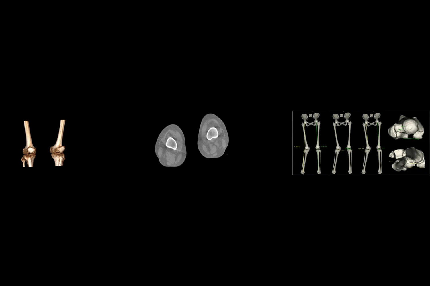
Reconstruction software combines the projections into volumetric data. Coronal, sagittal, and axial slices provide different views through the knee, allowing the joint region to be examined beyond a single projection.
3. Define the region and calculate local spacing
The analysis method defines the joint region and estimates spacing at multiple locations. Some methods calculate distances between reconstructed bone surfaces; others estimate JSW from the image data.
4. Display the measurements
Distances are associated with their anatomical locations to create a joint space map. Summary values may include a regional minimum and a compartment average. The minimum describes the narrowest measured location in the defined region; the average summarizes that region; the map shows how spacing varies within it. Colors encode distance according to the accompanying scale, rather than directly grading cartilage damage.
Turmezei and colleagues’ quantitative knee joint space mapping study demonstrates one research implementation of surface-based measurement and display. It illustrates the principle of preserving spatial information alongside numerical results.
What Does a 3D Joint Space Map Add to Osteoarthritis Assessment?
A joint space map connects a measurement to a place in the knee. Its value is clearest when location, distribution, and alignment are considered separately.
Locate the narrowing
A small minimum JSW value identifies a narrow measured gap. Mapping shows where that value occurs within the assessed compartment, so the corresponding anatomy can be reviewed on reconstructed slices.
Describe the distribution
A compartment average summarizes many locations. Two illustrative measurement maps could have similar averages while one contains a small area of narrowing and the other a broader distribution of lower values. Keeping the map and summary values together preserves that distinction.
Relate local findings to alignment
Alignment measurements add geometrical context. Joint line convergence angle describes the relative orientation of the femoral and tibial joint lines, while hip-knee-ankle alignment relates the knee to the limb’s overall axis. Where the appropriate scan coverage and measurements are available, these can be reviewed alongside compartment narrowing.
Our published knee measurement workflow describes medial and lateral JSW, joint line convergence angle, and joint-gap heat maps. These outputs illustrate how spacing and joint orientation can be presented together.
Weight-Bearing 3D Imaging vs. Standing X-Ray and Knee MRI
Standing radiography, weight-bearing 3D imaging, and MRI provide different views of knee structure. The comparison below separates the main image information from its contribution to assessment.
| Examination | Format/ loading | Main information | Role in assessment |
|---|---|---|---|
| Standing radiography | 2D projection under load | Projected joint space and bone changes | Overview of narrowing and alignment; positioning affects the projection |
| Weight-bearing 3D imaging | Reconstructed volume under a defined standing load | Bone anatomy and regional bone-to-bone spacing | Localizes narrowing and supports cross-sectional review |
| Knee MRI | Multiplanar imaging; routinely supine | Cartilage, menisci, ligaments, and bone marrow | Evaluates tissue-specific abnormalities |
Standing X-rays provide an established view of joint space narrowing and bone changes. Weight-bearing 3D imaging adds spatial localization and cross-sectional review under load. MRI provides more detailed assessment of cartilage, menisci, ligaments, and other tissues; the ACR and RSNA knee MRI guide describes these applications.
The examinations therefore provide complementary information. Regional bone-to-bone spacing and tissue-specific abnormalities are different findings, and a 3D spacing map does not replace MRI assessment of a meniscal tear or cartilage lesion.
Radiography and X-ray-based 3D imaging use ionizing radiation; MRI does not. Dose comparisons between X-ray examinations require the actual protocols, including scan coverage and acquisition settings.
How WR-3D Supports Weight-Bearing Knee Imaging
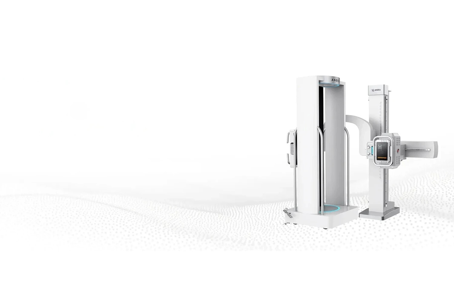
Our WR-3D weight-bearing imaging system combines standing volumetric acquisition with multiplanar reconstruction. Coronal, sagittal, and axial views allow the knee to be examined in cross-section, while full lower-limb imaging extends review to alignment across the limb.
The system uses a fixed X-ray tube and flat-panel detector while a motorized platform rotates the standing patient. This is the WR-3D acquisition design; maintaining the prescribed posture during rotation supports reconstruction quality.
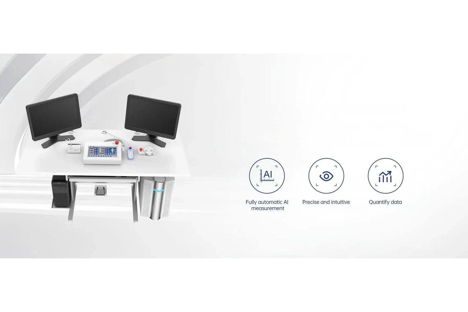
Relevant optional measurement packages provide AI-assisted quantification. Published knee outputs include medial and lateral JSW, joint line convergence angle, and a joint-gap heat map. These combine numerical measurements with location and orientation information for clinician review. WR-3D also supports 2D radiography and stitching workflows.
A published deployment example is the WR-3D installation at Cotabato Regional and Medical Center in the Philippines. The announcement documents installation and a live demonstration of standing imaging. It illustrates deployment in a hospital setting, rather than patient-level evidence of improved knee osteoarthritis outcomes.
Frequently Asked Questions
Does 3D joint space width measure cartilage thickness?
No. It measures or estimates the separation between opposing bone surfaces. Cartilage and meniscal changes can influence that separation. MRI provides tissue-specific information when direct assessment of cartilage or menisci is needed.
What do the colors on a joint space map mean?
They represent measured distances according to the map’s numerical legend. The same color can represent different distances on maps with different scales, so color alone is not a universal severity grade.
How does 3D assessment differ from a standing X-ray?
Both can examine the knee under load. A radiograph represents the anatomy as a projection; a volumetric examination supports cross-sectional viewing and spatial mapping of regional spacing.
Can 3D JSW support follow-up of knee osteoarthritis?
It can support structural comparisons when the acquisition and analysis methods are consistent and changes are assessed against measurement variability. It does not, by itself, establish the cause of symptoms or predict an individual’s future disease course.
How should patients prepare for weight-bearing 3D knee imaging?
For examinations using our WR-3D system, patients should follow the imaging department’s preparation instructions and wear clothing that allows comfortable positioning. Metal objects around the knees may need to be removed. Patients should also tell the team about mobility difficulties or a possible pregnancy before the examination so that suitability and positioning can be assessed.
Conclusion
Weight-bearing 3D imaging adds spatial detail to knee osteoarthritis assessment by showing where joint-space narrowing occurs under load and how it relates to alignment. Joint-space maps should be interpreted alongside reconstructed images and the clinical findings; they do not directly measure cartilage thickness or replace tissue-specific MRI assessment.
Our WR-3D system brings standing volumetric acquisition and multiplanar review into the orthopedic imaging workflow. If your department is evaluating this approach,contact our Angell team to discuss patient positioning, scan coverage, available measurement packages, and installation requirements.