
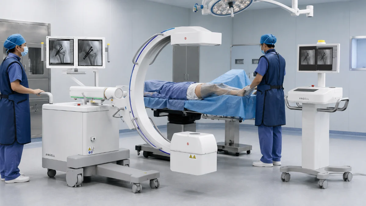
A C-arm is a mobile X-ray imaging system named after the curved structure that connects its X-ray tube and detector. The arc swings around a patient on an operating table, giving surgeons a live video feed — fluoroscopy — so they can watch instruments, implants, and anatomy in real time without repositioning the patient. Orthopedic, vascular, urological, and trauma teams all rely on C-arms for that intraoperative guidance.
At Angell Technology, our engineering team has worked across the DR imaging chain — flat-panel detectors, X-ray tubes, high-frequency generators, and acquisition software — for more than two decades. Our materials also document Angell’s participation in drafting the industry standard for dynamic digital X-ray imaging. In this guide, we explain how C-arms work, what they are used for, and what separates one system from another.
What Is a C-Arm Machine?
Why Is It Called a C-Arm?
Picture a large letter C lying on its side: the X-ray tube sits at one tip and the flat-panel detector at the other. That rigid curve keeps both components aimed at the same point on the patient no matter how the arm rotates, tilts, or slides — so a surgeon can move from a head-on (AP) view to a side (lateral) view by repositioning the C-arm rather than repositioning the patient. The range of that movement — orbital rotation, angulation, vertical lift, horizontal slide, and the open space inside the C — determines which projections the system can reach around a given operating table.
C-Arm Machine vs Fluoroscopy
A C-arm is the equipment; fluoroscopy is the imaging technique. The U.S. Food and Drug Administration describes fluoroscopy as a continuous X-ray image displayed on a monitor so clinicians can observe anatomy, an instrument, or contrast material in motion. Some C-arms also capture single radiographic frames.
That distinction matters when writing a purchase request. A department that asks for “fluoroscopy” could mean a mobile surgical C-arm, a fixed interventional suite, or a radiography/fluoroscopy room — three different equipment categories with different budgets and clinical workflows.
How Does a C-Arm Machine Work?
The imaging chain, step by step. When a surgeon steps on the foot switch, the X-ray tube fires a controlled burst of radiation through the patient. Dense structures — bone, metal implants, contrast dye — absorb most of that energy and appear bright on the monitor. Soft tissue lets more radiation through and appears as shades of gray. Air passes nearly everything and looks dark. The flat-panel detector on the opposite side of the C captures whatever radiation makes it through and converts it into a digital image in a fraction of a second.
Two modes: live video and still capture. In fluoroscopy mode, the tube fires repeated low-dose pulses at a rate set by the protocol and system configuration, producing a continuous video stream. That lets the surgical team watch a guidewire advance into a vessel or a fracture reduce under traction in real time. When a sharper record is needed, the system switches to radiographic mode: a single exposure that captures a still frame for documentation or assessment.
Changing the angle without moving the patient. Because the tube and detector are locked to the same C-shaped structure, the entire imaging axis pivots as one unit. During a hip-screw fixation, for example, the surgeon checks the frontal view to confirm alignment, then repositions the C-arm for a lateral view to judge screw depth — all while preserving the sterile workflow.
Why the full chain matters. The image a surgeon sees is shaped by every link in that chain: the tube’s focal-spot size, the generator’s output stability, the detector’s active area and dynamic range, and the software that converts raw signal into a viewable image. Because our R&D covers the detector, generator, tube, mechanical platform, and acquisition software, the imaging chain can be tuned as one system instead of being treated as isolated components.
What Are C-Arms Used For?
C-arms are most closely associated with orthopedic surgery, but fluoroscopic guidance supports a much broader range of interventions. FDA examples include fracture treatment, joint replacement, catheter manipulation, stent placement, and angiography.
| Application | What the C-arm helps clinicians observe |
|---|---|
| Orthopedic surgery | Fracture reduction, fixation hardware, joint alignment, and implant position. During a femoral nail procedure, the AP view shows overall alignment while the lateral confirms the nail is not protruding through the cortex. |
| Vascular and interventional procedures | Guidewires, catheters, stents, and contrast flow in selected interventional workflows. High-volume coronary or neurovascular work is usually handled in a dedicated fixed suite rather than a general mobile C-arm workflow. |
| Urology | Access paths, instruments, and urinary stents. In percutaneous nephrolithotomy, fluoroscopy guides the access needle into the kidney’s collecting system under real-time imaging. |
| Emergency and trauma care | Imaging support when a patient cannot be transferred to a fixed X-ray room, especially in trauma OR workflows. For bedside emergency observation outside the OR, mobile dynamic DR may be the better fit. |
Types of C-Arm Machines
Mini C-Arms vs Full-Size Mobile C-Arms
Mini C-arms are usually built for extremity imaging — hand, wrist, foot, and ankle — where the field of view and generator output can be smaller. Full-size mobile C-arms are built for larger anatomy such as the spine, hip, pelvis, and abdomen, so buyers should compare detector coverage, generator power, tube heat capacity, gantry opening, and room fit rather than relying on the marketing label alone.
Fixed C-Arms vs Mobile C-Arms
A fixed C-arm is mounted in a dedicated interventional suite, often integrated with a specialized table, multiple monitors, and hemodynamic equipment. It stays in one room and handles high-volume catheterization or neuro-interventional work. A mobile C-arm travels between ORs on wheels and works with whatever surgical table is already in place. The two serve fundamentally different workflows.
2D Fluoroscopy vs 3D-Capable C-Arms
A standard mobile C-arm produces two-dimensional views from selected projections. A 3D-capable C-arm acquires images over a motorized rotational sweep and reconstructs them into a volumetric (cone-beam CT) dataset. That capability requires motorized rotation, calibration geometry, reconstruction software, and regulatory clearance. A flat-panel detector is necessary but not sufficient: 3D is a system-level capability, not a detector feature alone. In Angell’s C-arm roadmap, intraoperative 3D is treated as a future system-level direction linked with high-precision gantry motion and WR-3D software know-how, not as a standard feature of every mobile C-arm.
FPD C-Arm vs Image Intensifier C-Arm
An image intensifier (II) converts X-rays into light inside a vacuum tube, producing a circular image field with geometric distortion toward the edges. A flat-panel detector (FPD) uses a flat detector array to produce a rectangular image with more uniform geometry in a slimmer housing. Around an operating table, that slimmer profile gives the surgical team more room for instruments and lateral positioning.
FPDs also offer wider dynamic range and greater stability over time. Some II-based systems still deliver higher spatial resolution in certain magnification modes and cost less upfront. What matters most is the complete imaging chain — generator, detector, processing, and protocol — not the detector label alone.
Main Parts of a C-Arm System
| Component | Role | Why it matters |
|---|---|---|
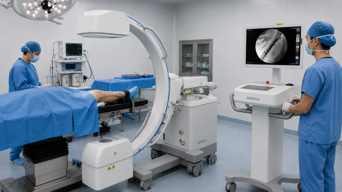
| X-ray tube and generator | Produces and controls the X-ray beam | Generator kW and tube heat capacity determine how long the system can image before cooling — critical in lengthy cases |
| Collimator | Limits the radiation field to the area of interest | Tighter collimation reduces patient dose and improves contrast by cutting scatter |
| Flat-panel detector (or image intensifier) | Converts incoming X-rays into a digital image | Pixel pitch, active area, and dynamic range directly affect sharpness and field coverage |
| C-shaped gantry | Holds tube and detector in alignment; rotates around the patient | Orbital range and open space inside the C determine reachable projections and surgical-team clearance |
| Mobile base and brakes | Supports transport and locks the system in position | Base width and turning radius decide whether the unit passes through OR doors |
| Workstation and monitors | Process, display, and store images | Monitor position and processing speed affect the surgeon’s line of sight and workflow |
No single number tells the whole story. Tube output, detector design, software, and workflow have to work together. When detector design, tube performance, generator output, and acquisition software are developed and validated as one imaging chain, the system is easier to tune for consistent clinical images — which is why our R&D spans the full chain from detector design to acquisition software.
When Is a C-Arm the Right Imaging System?
Not every procedure that needs X-ray guidance needs a C-arm. Several equipment categories provide dynamic or mobile imaging, but each addresses a different clinical question.
| Clinical need | Equipment category | Angell system category |
|---|---|---|
| Real-time guidance during surgery — fracture fixation, screw placement, orthopedic intraoperative guidance | Mobile C-arm | HUA II Series (ALC280 Series) Mobile Dynamic FPD C-arm |
| Bedside or emergency imaging with dynamic observation — PICC guidance and verification, intubation checks, and lung/diaphragm motion observation | Mobile Dynamic DR | Lingxi Series — our mobile dynamic DR for bedside dynamic imaging |
| Multi-position radiography and fluoroscopy in a fixed room | UC-ARM Dynamic DR | DTP580 Series |
| Standing, weight-bearing 3D assessment of spine, hips, knees, and ankles | Weight-Bearing 3D Imaging | WR-3D with optional AI automatic measurement |
A mobile C-arm is the right starting point when the imaging question arises during the procedure, the clinical team needs to change projection around the table, and the answer must appear on-screen in real time.
How We Design Mobile Dynamic FPD C-Arms: HUA II Series
Modular design for different room layouts. In our C-arm materials, the HUA II Series (ALC280 Series) is positioned as the first split/all-in-one dynamic flat-panel mobile C-arm. It can switch between an all-in-one configuration and a split configuration with a separate monitor cart to match room layout, sightlines, teaching needs, and transport routes.
Six low-dose safety features. In our C-arm design, dose management is built around 200 kHz high-frequency high-voltage generation, mA/ms separate exposure settings, APR organ-program exposure, DAP dose display and monitoring, dual laser positioning, and three exposure modes including wired foot-switch, wired hand-switch, and wireless remote exposure up to 25 m. ABS and IDC support image consistency and exposure control during imaging.
Dynamic imaging and data handling. The system’s dynamic flat-panel detector supports fluoroscopy and still-frame capture, helping clinicians adjust implant position during surgery and assess implant stability after fixation or joint movement. Export formats and PACS/DICOM integration should be confirmed according to the selected configuration and destination-market requirements.
Looking ahead, our C-arm roadmap treats larger detector coverage, intraoperative 3D imaging, spinal deformity measurement, and dual-energy flat-panel exploration as future directions for more complex orthopedic scenarios.
How to Choose a C-Arm Machine
A useful configuration brief starts with four questions:
Clinical requirements. Record the procedure mix, anatomical regions, patient size range, required projections, and expected weekly case volume. The detector must cover the anatomy, implants, and instruments in a single field.
Generator and tube performance. Match generator kW, tube heat capacity, and cooling rate to patient thickness, procedure duration, and daily workload. For high-volume ORs, request documented heat-capacity and cooling-curve data.
Physical fit in the OR. Map the C-arm’s gantry opening, orbital travel, base width, and monitor position against the surgical table, anesthesia zone, sterile field, doorways, and cable routes. An operating room can look spacious on a floor plan and still force a positioning compromise — if the detector cannot reach the required lateral view without entering the anesthesia zone, the system will interrupt the workflow even though it fits through the door.
Image handling, service, and compliance. Confirm capture modes, dose display, export formats, and whether PACS/DICOM integration is included in the selected configuration. Then close the loop on installation, acceptance testing, detector calibration, operator training, preventive maintenance, and regulatory clearance in the destination market.
Radiation Safety and Dose Management
Fluoroscopy uses ionizing radiation. Dose depends on procedure length, patient size, anatomy, projection angle, pulse rate, collimation, and operator technique — not on the detector type alone. A flat-panel detector does not automatically mean “low dose.” The FDA recommends the lowest acceptable exposure for the shortest necessary time; the IAEA adds pulsed fluoroscopy at the lowest suitable pulse rate, tight collimation, last-image hold, and routine quality-assurance testing. Equipment features support dose optimization, but protocol design, staff training, and facility QA complete the picture.
Frequently Asked Questions About C-Arm Machines
Is a C-arm the same as fluoroscopy?
No. A C-arm is the hardware — the source-detector system on a mobile base. Fluoroscopy is the imaging technique that produces continuous X-ray video. Fixed interventional suites also provide fluoroscopy; they just use a different equipment form factor.
What is the difference between a C-arm and a standard X-ray machine?
A mobile C-arm provides live imaging during a procedure and can change projection angle on the fly. A general radiography system is built for diagnostic still images in a radiology room or at the bedside. A C-arm is designed specifically for intraoperative guidance around a surgical table.
What is the difference between a mini and full-size C-arm?
Size, coverage, and power. A mini C-arm images extremities with a small detector and a compact frame. A full-size C-arm covers broader anatomy with a larger detector, higher generator output, and greater tube heat capacity. Choose based on the anatomy and projections the department needs most often.
Does every C-arm produce 3D images?
No. 3D or cone-beam CT imaging requires a motorized rotational sweep, reconstruction software, calibration geometry, and regulatory clearance. Most mobile C-arms produce 2D fluoroscopy and radiography only.
What should hospitals prepare before requesting a quotation?
Procedure mix, anatomical coverage, patient size range, required projections, estimated weekly case volume, OR floor plan with door widths, table specifications, PACS/DICOM environment, destination-market regulatory requirements, and service expectations.
Conclusion
A C-arm brings live X-ray guidance to the procedure table, but its clinical value comes from the fit between the imaging chain, gantry movement, room geometry, exposure controls, and the planned case mix. Not every imaging need calls for a C-arm — mobile dynamic DR, UC-arm radiography, and weight-bearing 3D each answer different clinical questions.
Send us the procedure mix, OR floor plan, table dimensions, expected case volume, and destination market. Our technical team will prepare a configuration brief for the HUA II Series or recommend the right system category for the workflow.