
Mobile Dynamic FPD C-arm medical imaging solutions

Mobile Dynamic FPD C-arm

Product Categories

Product Category

Mobile Dynamic FPD C-Arms

Flat panel detector C-arms built for imaging inside the operating room.

What is a mobile dynamic FPD C-arm?

A mobile C-arm is a wheeled X-ray system whose C-shaped arm carries an X-ray source at one end and a detector at the other. Because the arm orbits around the patient without the patient being moved, the surgical team can see live X-ray images of anatomy and instruments during a procedure, and capture still images when a position needs to be documented.

"Dynamic FPD" describes how the image is formed. Instead of the image intensifier tube used by earlier C-arm generations, a dynamic flat panel detector converts X-rays into a digital signal directly on a flat sensor, at video frame rates. The result is a flat, rectangular image field with none of the geometric distortion and edge darkening inherent to a curved intensifier input surface — and a housing thin enough to leave more working room around the table.

Angell has developed its own flat panel detector, X-ray tube and high-voltage generator since 2002, and applies that detector technology to the C-arm systems in this category.

Where it is used

The operating room and interventional suite — not the radiology examination room. Imaging happens while the procedure is under way.

What it replaces

Image intensifier C-arms, which remain widely installed but lose image quality as the intensifier tube ages.

How to size it

Match the detector field of view, arm depth and free space to the procedures you run most often, then check the dose-management features.

Category scope

Mobile systems only. For fixed-installation radiography, see the ceiling-mounted and floor-mounted categories.

In this category

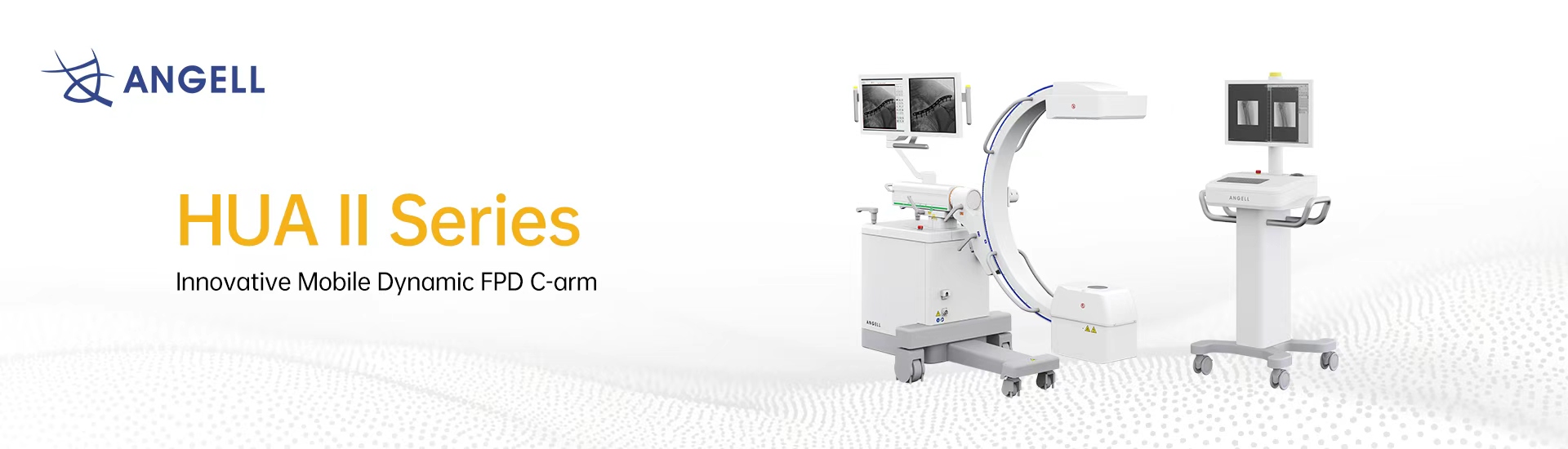
HUA II Series

Innovative Mobile Dynamic FPD C-arm

Angell's mobile dynamic FPD C-arm platform, equipped with a dynamic flat panel detector and a set of low-dose safety designs for use throughout a procedure. Full feature breakdown, configurations and specifications are on the product page.


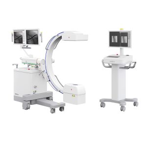
HUA II Series mobile dynamic FPD C-arm by Angell Technology

Flat panel detector C-arm vs. image intensifier C-arm

Both designs produce live X-ray images. The difference is the detector at the receiving end of the arm, and that difference shapes image geometry, the shape of the field you see, and how much room the arm leaves you at the table.

Flat panel detector (FPD) C-armImage intensifier (I.I.) C-arm
Detection principleX-rays are converted on a flat digital sensor and read out directly at video rate.X-rays strike a curved input screen; the resulting light image is intensified through a vacuum tube, then picked up by a camera.
Image field shapeRectangular — the full sensor area is usable.Circular, so the corners of the display carry no image.
Geometric distortionFlat sensor surface, so no pincushion distortion and no S-distortion from ambient magnetic fields.Curved input surface and electron optics introduce pincushion and orientation-dependent distortion.
Brightness uniformityUniform response across the field.Vignetting — the image tends to fall off toward the edges.
Performance over service lifeNo intensifier tube to age; output stays stable.Intensifier gain degrades with age, which typically pushes dose up to hold image quality.
Form factor at the tableA slim detector housing, leaving more free space around the patient.A deep, bulky intensifier housing on the receiving end of the arm.

Comparison of the two detector architectures in general terms. Field of view, resolution and dose behaviour depend on the individual system — see the product page for the specifications of a given model.

Where these systems are used

A mobile C-arm is chosen for the procedure it has to follow. These are the four settings that most often drive the specification.

01

Orthopedic surgery

Fracture reduction, fixation and joint work, where implant and instrument position is confirmed live and re-checked before closing.

02

Vascular procedures

Guidewire, catheter and device navigation, where contrast runs have to be followed continuously and replayed.

03

Urology

Stone and access work, where the arm must move between projections around a patient who stays in position.

04

Emergency & trauma

Unplanned cases, where the system has to be moved in, positioned and imaging within a short setup window.

Choosing the right category

Systems in this category image during a procedure, with the C-arm brought to the table. If instead you need imaging before or after surgery — standing alignment assessment, planning, follow-up — that is a different class of system, and the categories below are the right starting point.

Talk to us about your operating room

Tell us the procedures you run and we will come back with the configuration, specifications and pricing that fit.

Get a Quote