
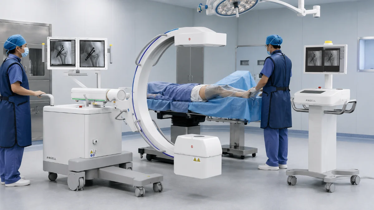
What Is a C-arm Machine — and What Does It Actually Do in Surgery?
A C-arm is a wheeled X-ray system whose C-shaped arm carries an X-ray source at one end and a detector at the other. The arm orbits around the patient without moving the patient, allowing the surgical team to view live X-ray images of the anatomy and instruments. At the same time, a procedure is underway — and it captures still frames when a position needs to be documented.
That real-time loop between imaging and action is what separates a C-arm from a standard X-ray room. A conventional radiography system captures a single exposure, then the clinician reviews the result and decides the next step. A C-arm closes that gap: the image updates continuously, the surgeon adjusts in response, and confirmation happens before the incision is closed rather than after.
Since 2002, we have worked in digital medical imaging. In our experience, hospitals get more value from a C-arm when the system is chosen around real procedure mix, operating room layout, and daily staffing habits. This article walks through the technology, clinical scenarios, and decision points that matter in practice.
Inside the Machine: Core Components of a C-arm
A C-arm is not a single device; it is an imaging chain assembled from several interdependent parts. Understanding each one helps explain why two systems with similar-looking spec sheets can behave very differently in daily use.
X-ray tube. The radiation source. Tube design affects maximum output power, heat capacity, and how many consecutive exposures the system can sustain before it needs a cooling pause — a factor that matters in long orthopedic or vascular cases.
High-voltage generator. Converts input power into the kilovoltage that drives the tube. Generator quality determines how precisely exposure parameters can be controlled, which feeds directly into dose management.
Detector. Sits opposite the tube and captures the X-ray signal. This component has a major influence on image quality, field shape, dose-control behavior, and the working space around the patient.
Collimator. Shapes the X-ray beam before it reaches the patient, restricting exposure to the region of interest and reducing unnecessary dose to surrounding tissue.
C-shaped gantry and mechanical frame. The structure that holds the tube and detector in alignment while allowing the arm to rotate, tilt, and slide around the patient. Arm depth and free space determine how much working room the surgical team has at the table.
Image processing workstation. Converts raw detector data into the fluoroscopic images displayed on the monitor, and handles storage, export, and post-processing.
We develop and manufacture several of these core components in-house — including X-ray tubes, high-voltage generators, flat panel detectors, collimators, and mechanical assemblies. Controlling these parts of the chain is what lets us tune the system as a whole.
Flat Panel Detector vs. Image Intensifier: Why the Detector Choice Matters
A major technical distinction in the C-arm market is the detector. Image intensifier (I.I.) systems use an image intensifier tube — a vacuum tube with a curved input surface that converts X-rays into visible light, then amplifies the signal through electron optics before a camera picks it up. FPD systems use a dynamic flat panel detector (FPD) — a flat digital sensor that converts X-rays directly into a digital signal at video frame rates.
The practical differences go well beyond image quality:
| Factor | Flat Panel Detector (FPD) C-arm | Image Intensifier (I.I.) C-arm |
|---|---|---|
| Detection principle | X-rays convert on a flat digital sensor and are read out directly at video rate | X-rays strike a curved input screen; the light image is intensified through a vacuum tube, then picked up by a camera |
| Image field shape | Rectangular — the full sensor area is usable | Circular — corners of the display carry no image |
| Geometric distortion | Helps avoid pincushion distortion and S-distortion associated with curved image intensifier designs | Curved input surface and electron optics introduce pincushion and orientation-dependent distortion |
| Brightness uniformity | Uniform response across the field | Vignetting — image brightness falls off toward the edges |
| Performance over service life | No image intensifier tube in the detector chain | Aging intensifier performance may require exposure adjustment to maintain image quality |
| Form factor at the table | Slim detector housing leaves more free space around the patient | Deep, bulky intensifier housing on the receiving end of the arm |
The service-life point deserves emphasis because it is often invisible during a product demo. An image intensifier can perform well when new, yet intensifier tube performance can change with age. Long-term evaluation should look at image consistency, exposure-control behavior, and maintenance expectations, especially when the same room supports repeated fluoroscopy cases.
Where C-arm Systems Are Used: Four Scenarios, Four Different Demands
Orthopedic Surgery
Orthopedic surgery is one of the most common application areas for mobile C-arm systems. The surgeon reduces the fracture, places hardware, and confirms alignment under live fluoroscopy — often re-checking from multiple angles before closing. In a typical long-bone fixation, the C-arm may be repositioned several times to verify screw length, plate alignment, and joint congruence. What matters here is positioning flexibility and image stability: the arm has to reach anteroposterior and lateral views quickly, and the image needs to stay consistent across repeated exposures.
Urology
Lithotripsy, ureteroscopic stone removal, percutaneous renal access, and ureteral stent placement all rely on continuous fluoroscopic guidance. The C-arm confirms guidewire position, tracks instrument advancement, and verifies stent deployment — all without moving the patient. Some urology procedures may include extended fluoroscopy sequences, so dose management and detector stability need review across longer runs as well as short spot checks.
Vascular Procedures
Guidewire and catheter navigation, stent deployment, and contrast-enhanced vessel visualization require a C-arm that can follow device movement in real time. Contrast runs need to be watched continuously and often replayed frame by frame to confirm device position. Image processing speed and the ability to capture and store sequences mid-procedure become deciding factors.
Emergency and Trauma
In emergency and trauma cases, the value of a mobile C-arm is fast confirmation at the procedure table. The team may need to check fracture reduction, hardware position, or instrument placement while patient preparation and surgical treatment are happening at the same time. A suitable system should move into position smoothly, reach the needed view with minimal repositioning, and provide a clear image quickly enough for the surgeon to make the next decision.
How a Modular Dynamic FPD C-arm Addresses These Scenarios
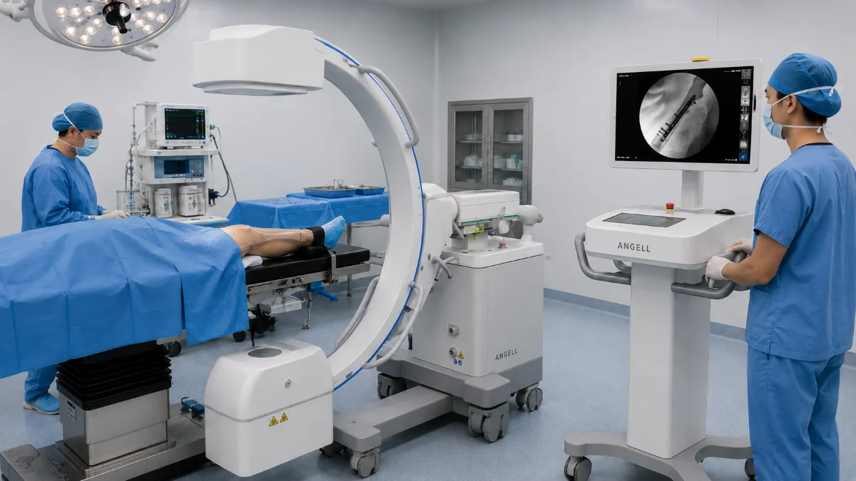
Modular Configuration: All-in-One or Split
Our HUA II Series is an industry-first modular dynamic FPD mobile C-arm that converts between all-in-one and split configurations. In all-in-one use, the monitor, controls, and processing stay on the mobile base, which can reduce the number of separate pieces the team needs to position. In split use, the monitor and workstation can be separated from the C-arm body, giving staff more freedom to arrange sightlines and movement around the table.
Monitor posture is part of that workflow. In all-in-one use, the three-axis articulated monitor arm can be adjusted to reduce screen obstruction from the C-shaped gantry, so the operator can check the image from the working position. In split use, the monitor cart can be placed where an assistant or teaching observer needs a clear view, which helps when one operating room supports more than one procedure type.
Six Low-Dose Safety Designs
On the HUA II Series, radiation management comes from six coordinated designs:
Auto Exposure adjusts parameters automatically based on the anatomy being imaged. Automatic Program Response (APR) loads pre-set protocols matched to procedure type. Laser positioning marks the beam center on the patient’s body so the operator can align without a test exposure. Automatic Brightness Stabilization (ABS) and Instant Dose Control (IDC) work together to hold image brightness steady while minimizing dose fluctuation during live fluoroscopy. Dose Area Product (DAP) display and analytics help the team monitor and review dose-related information during imaging workflows. Remote exposure allows the operator to trigger imaging from outside the direct radiation field.
Each of these addresses a different moment in the procedure. Laser positioning matters before the first exposure. ABS and IDC matter during long fluoroscopy runs. DAP analytics matter when the team reviews total dose after the case. The combined workflow matters more than any single feature.
Imaging and Export Workflow
Our HUA II Series supports multiple image capture modes, multi-frame imaging acquisition, and all-format image export. In practice, this supports confirmation images, fluoroscopy sequences, documentation, review, and archiving workflows without interrupting the procedure.
How to Choose the Right C-arm for Your Operating Room
The strongest selection process starts from the procedures performed before moving to the equipment catalog.
Match Detector Field of View and Arm Depth to Your Procedures
A C-arm used primarily for distal extremity work (wrist, ankle) does not need the same field of view as one used for pelvic or spinal procedures. Arm depth — the distance from the open side of the C to the center of the image field — determines whether the system can reach across a standard operating table without interference. Measure your tables, check the arm specifications, and test clearance with the patient positioning you actually use.
Evaluate Dose Management as a System, Not a Checkbox
A low-dose claim is more useful when it is connected to clear exposure-control mechanisms. Ask how the system manages exposure during continuous fluoroscopy, whether dose-related information can be reviewed, and whether remote exposure is supported. These details influence radiation management across repeated clinical use and can affect cumulative staff and patient exposure during fluoroscopy-guided work.
Consider the Room, Not Just the Machine
Even a capable C-arm can create workflow friction if it does not fit the room layout. Operating room dimensions, anesthesia equipment, staff working positions, and monitor visibility all affect whether a system integrates smoothly into daily procedures.
When a Mobile C-arm Is Not the Right System
A mobile C-arm is mainly designed for intraoperative imaging, with real-time guidance brought directly to the procedure table. Its role is different from a radiology examination room system, where workflow centers on scheduled diagnostic examinations and fixed room geometry.
If the clinical need is pre-operative or post-operative imaging, standing alignment assessment, full-spine or full-limb evaluation, or general radiography, the starting point is a different class of system. Ceiling-mounted dynamic DR systems, floor-mounted radiographic systems, and weight-bearing 3D imaging systems are designed around examination-room workflows rather than mobile intraoperative guidance.
A related category is the UC-arm. A UC-arm dynamic DR system (such as our DTP580 Series) may look related because the tube and detector sit at opposite ends of a rigid arm. Its clinical role is different: the UC-arm is a fixed-installation system for radiology rooms that handle multi-position examinations. It supports specialized projections like Waters view, patella axial view, calcaneal axial view, and cross-table lateral view by rotating the arm and detector. These views are enabled by fixed-arm geometry and detector rotation, while a mobile C-arm is chosen for intraoperative access at the procedure table. Separating the two categories helps hospitals match the right equipment to the right room and workflow.
Manufacturer Capability Behind the Equipment
A C-arm will be in clinical service for years. During that time, hospitals need consistent technical support, spare parts availability, software updates, operator training, and workflow support. Manufacturer capability should be evaluated together with product specifications.
We are one of the few manufacturers with technology covering X-ray detectors, X-ray tubes, and X-ray generators. This capability helps us connect component-level performance with complete system design, image acquisition software, service training, and long-term technical support.
By 2025, our installed base had reached 30,000 units across more than 100 countries, supported by a team of nearly 600 employees. Our product categories span mobile dynamic FPD C-arm, mobile DR, ceiling-mounted dynamic DR, UC-arm dynamic DR, digital R/F table systems, floor-mounted radiographic systems, digital mammography, weight-bearing 3D imaging, and core DR components.
FAQs
What is a C-arm machine used for?
A C-arm provides real-time X-ray imaging during surgical and interventional procedures, including orthopedic fixation, urology interventions, vascular catheterization, and emergency trauma cases. Its main role is imaging while the procedure is underway.
What is the difference between an FPD C-arm and an image intensifier C-arm?
An FPD C-arm uses a flat digital sensor that provides a rectangular image field and helps reduce geometric distortion. An image intensifier C-arm uses a vacuum tube with a curved input surface, producing a circular image field with inherent geometric distortion, and its performance can change as the intensifier tube ages, which may require exposure adjustment to maintain image quality over time.
How do I know which C-arm is right for my hospital?
Start with the procedures you run most often. Match the detector field of view and arm depth to those cases, check that the system fits your operating room dimensions, and evaluate dose management features as a complete system rather than isolated line items on a specification sheet.
Can a C-arm replace a radiology room X-ray system?
No. A mobile C-arm is built for intraoperative imaging during procedures. Examination-room workflows, such as general radiography, standing alignment, and multi-position diagnostic projections, are better matched to fixed-installation systems such as ceiling-mounted DR, floor-mounted DR, or UC-arm dynamic DR.
A C-arm Is a Workflow Decision, Not a Specification Decision
The right C-arm is the one that fits the procedures your surgical team actually performs, the operating room they work in, and the dose management standards your hospital maintains. Technology matters — flat panel detectors, modular configurations, and integrated dose controls all contribute to clinical performance — but technology only delivers value when it is matched to the environment.
If you are evaluating a mobile dynamic FPD C-arm system, our technical team is available to review your procedure mix, operating room layout, and workflow requirements before recommending a configuration.