Product Category
Multipurpose Radiography & Fluoroscopy Tables for GI and Multidisciplinary Use
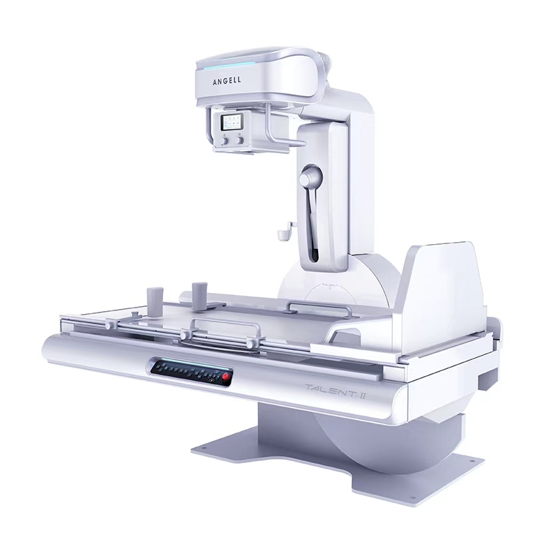
A digital R/F table system puts two examinations on one remote-controlled table: still radiography for structure, and live fluoroscopy for motion. A barium swallow, a contrast study and a routine abdominal view can all be completed in the same room, on the same patient position, without moving to a second machine.
R/F table systems from Angell
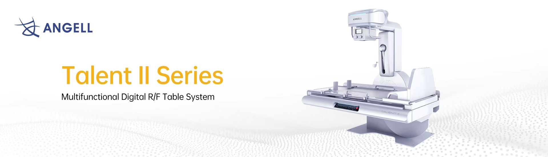
Multifunctional digital R/F table system
Angell's remote-controlled R/F table for departments that run radiography, fluoroscopy and contrast-enhanced examinations from a single room. Built around a dynamic flat panel detector developed in-house, it covers gastrointestinal studies, routine radiographic views and full-length stitched imaging, with a low-dose setup for paediatric and gynaecological patients.

Two modes, one table
The two modes answer different clinical questions. Understanding which one a department actually needs — and how often it needs both — is the first step in specifying a DR RF system.
Mode 01 — Radiography
A single high-dose-efficiency exposure records anatomy at rest. This is the mode used for chest, spine, abdomen and extremity views, and for full-length stitched images of the spine or lower limbs.
Mode 02 — Fluoroscopy
A continuous or pulsed low-dose beam shows anatomy as it moves — swallowing, peristalsis, contrast filling a cavity. The radiologist watches in real time and captures spot images only when the finding appears.
Where a fluoroscopy bucky table fits. If a department only images anatomy at rest, a radiographic room is enough. The moment a study depends on watching something happen — contrast moving, a joint articulating, a swallow completing — the room needs fluoroscopy, and an R/F table delivers both without a second installation.
Where R/F tables are used
An R/F table is rarely bought for a single specialty. Its value comes from the number of referring departments that can be served from the same suite.
Gastroenterology
Barium swallow, oesophagus, upper GI series and small-bowel follow-through depend on watching contrast move. This is the core workload of a gastrointestinal X-ray table and the reason most R/F suites exist.
Orthopaedics
Stitched full-spine and full-lower-limb images for alignment review, plus standard joint and extremity radiographs — captured on the same table used for contrast work earlier in the day.
Gynaecology
Hysterosalpingography needs live imaging of contrast filling and passing through the tubes. Dose control matters more here than in almost any other routine study.
Paediatrics
Children move, and every second of fluoroscopy counts. Pulsed acquisition, tight collimation and a removable grid keep exposure to the minimum the diagnosis allows.
Dose in fluoroscopy
Fluoroscopy is the only routine radiographic examination where the beam stays on while the clinician works. For paediatric and gynaecological patients, dose management is a specification question, not an operator habit.
Delivering the beam in pulses rather than continuously cuts exposure time while keeping the sequence readable.
Grids improve contrast in adults but add dose. Being able to remove one is what makes a room genuinely usable for small patients.
Restricting the field to the region of interest before the beam is on is the single most effective dose reduction available.
The cheapest dose saving is the exposure that never has to be repeated. Camera-assisted positioning and stored protocols reduce repeat rates.
Talk to an application specialist
Tell us the examinations you run and how often. If fluoroscopy is occasional, a different configuration may serve you better — we will say so.

Angell Technology established in 2002, is a global professional digital medical imaging equipment supplier.
Copyright © 2026 Angell Technology, All Right Reserved