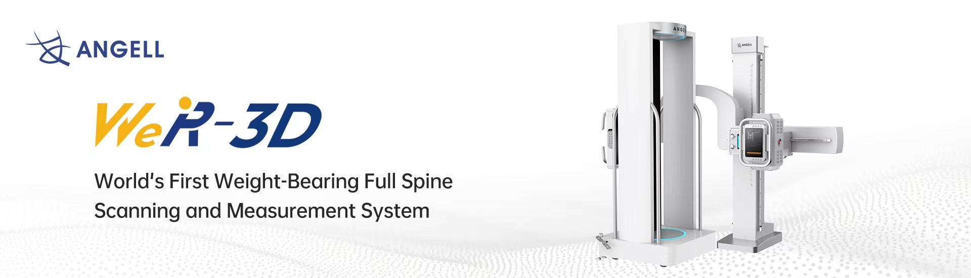
World’s First Weight-Bearing Full Spine Scanning and Measurement System
Imaging that scans the patient standing, under natural load — revealing the true alignment of the spine and lower limb that lying-down CT and MRI cannot show.
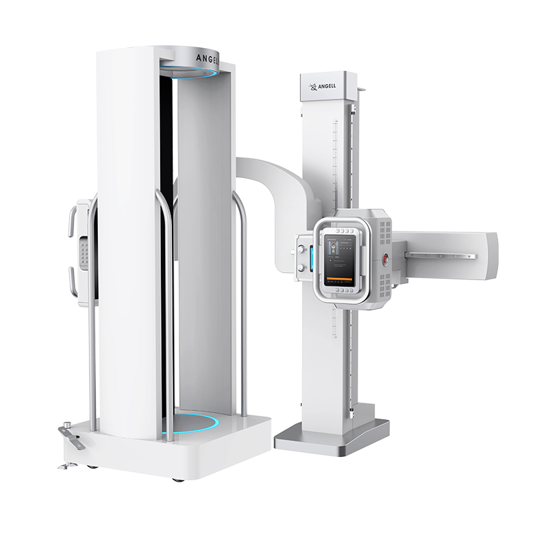
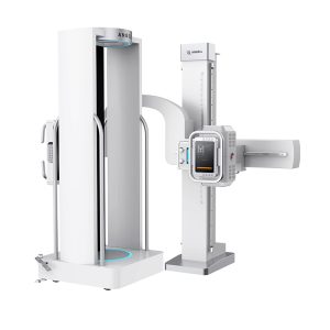
Angell's flagship weight-bearing 3D imaging system delivers whole-body volumetric imaging of the spine, hip, knee and ankle in the natural standing position — the world's first system of its kind, with AI-assisted measurement and 2D fluoroscopy on a single platform.
The skeleton behaves differently under gravity. Weight-bearing 3D imaging scans the patient standing, under natural physiological load, then reconstructs a full volumetric model of the bones and joints as they actually bear weight.
Conventional CT and MRI image the body lying down, with load removed — which can hide the very deformities and alignment shifts clinicians most need to see. By capturing the spine and lower limb in their functional posture, this category reveals the true biomechanical picture for diagnosis, surgical planning and follow-up. It complements CT and MRI rather than replacing them.
Each modality answers a different clinical question. This is where the category is uniquely positioned for orthopedic alignment and deformity.
| Weight-Bearing 3DStanding · loaded | Conventional CTSupine | MRISupine | |
|---|---|---|---|
| Patient position | Natural standing, weight-bearing | Lying (supine) | Lying (supine) |
| Skeletal load state | Under true physiological load | Unloaded | Unloaded |
| Reveals load-dependent alignment | Yes | No | No |
| Volumetric 3D reconstruction | Yes | Yes | Yes (soft tissue) |
| Full spine to ankle in one scan | Yes | Limited | Limited |
| Primary strength | Alignment, deformity & pre-op planning under load | Cross-sectional bone detail | Soft tissue & cartilage |
This category complements CT and MRI — it fills the gap they structurally cannot: the loaded, functional posture.
From full-spine balance to hindfoot alignment, weight-bearing 3D supports diagnosis, surgical planning and follow-up across the orthopedic pathway.
Scoliosis, sagittal balance and whole-spine alignment in the standing posture.
Dysplasia, pelvic tilt and the hip–spine relationship under natural load.
Osteoarthritis and varus / valgus alignment assessed while bearing weight.
Hindfoot alignment and instability in the loaded, functional position.
Mechanical-axis and leg-length mapping for osteotomy planning.
Request the WR-3D catalog and specifications, or talk to our team about clinical and installation requirements.

Angell Technology established in 2002, is a global professional digital medical imaging equipment supplier.
Copyright © 2026 Angell Technology, All Right Reserved